| Rabbit Anti-VEGF-C antibody |
| 产品应用(已验证) |
WB,IHC,FCM |
| 产品应用(可尝试) |
IF,ELISA |
| 推荐稀释比例 |
WB=1:500-2000,Elisa=1:5000-10000,IHC-P=1:100-500,IHC-F=1:100-500,Flow Cyt=1ug/Test,IF=1:100-500, |
| 研究领域 |
细胞生物,血管内皮细胞, |
| 标签 |
Array |
-
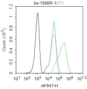
Blank control: HepG2.
Primary Antibody (green line): Rabbit Anti-VEGF-C antibody (bs-1586R)
Dilution: 1μg /10^6 cells;
Isotype Control Antibody (orange line): Rabbit IgG .
Secondary Antibody : Goat anti-rabbit IgG-AF647
Dilution: 1μg /test.
Protocol
The cells were fixed with 4% PFA (10min at room temperature)and then permeabilized with 0.1% PBST for 20 min at room temperature. The cells were then incubated in 5%BSA to block non-specific protein-protein interactions for 30 min at room temperature .Cells stained with Primary Antibody for 30 min at room temperature. The secondary antibody used for 40 min at room temperature. Acquisition of 20,000 events was performed.
-
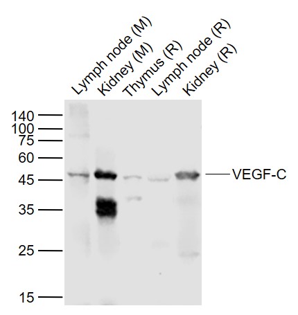
Sample:
Lane 1: Lymph node (Mouse) Lysate at 40 ug
Lane 2: Kidney (Mouse) Lysate at 40 ug
Lane 3: Thymus (Rat) Lysate at 40 ug
Lane 4: Lymph node (Rat) Lysate at 40 ug
Lane 5: Kidney (Rat) Lysate at 40 ug
Primary:
Anti-VEGF-C (bs-1586R) at 1/1000 dilution
Secondary: IRDye800CW Goat Anti-Rabbit IgG at 1/20000 dilution
Predicted band size: 46 kD
Observed band size: 46 kD
-
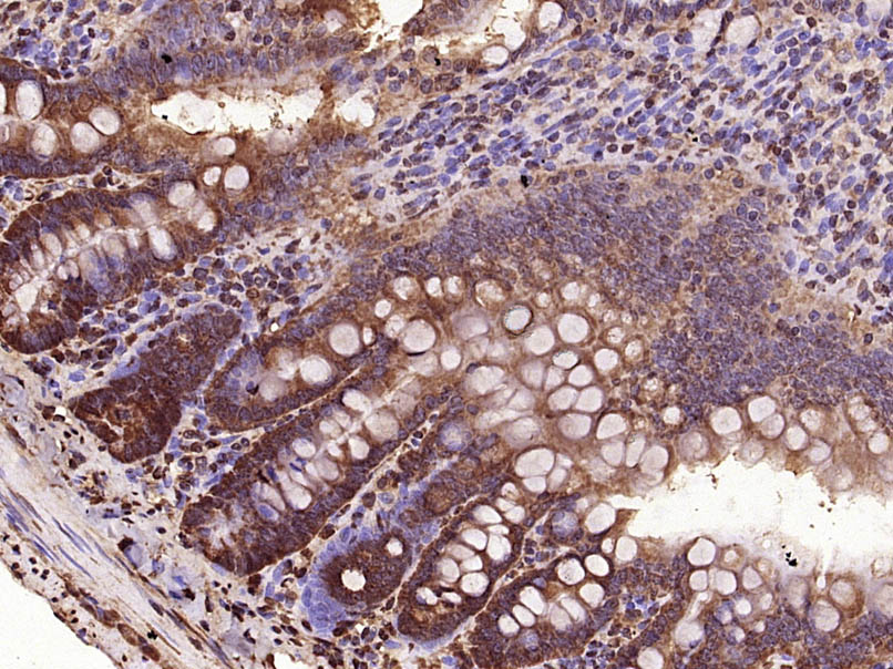
Paraformaldehyde-fixed, paraffin embedded (Rat small intestine); Antigen retrieval by microwave in sodium citrate buffer (pH6.0) ; Block endogenous peroxidase by 3% hydrogen peroxide for 30 minutes; Blocking buffer (3% BSA) at RT for 30min; Antibody incubation with (VEGF-C) Polyclonal Antibody, Unconjugated (bs-1586R) at 1:400 overnight at 4℃, followed by conjugation to the secondary antibody (labeled with HRP)and DAB staining.
RRID:AB_10856034
产品名称:Rabbit Anti-VEGF-C antibody
别名: Vascuoar endothelial growth factor-C; AW228853; Flt4 ligand; Flt4-L; VEGF2; VEGFC; VRP; VEGFC_HUMAN.
中文名称:血管内皮生长因子C型抗体
英文名称:Rabbit Anti-VEGF-C antibody
抗体来源: Rabbit
克隆类型:多克隆
细胞定位:分泌型蛋白
性 状:Liquid
亚 型:IgG
纯化方法:affinity purified by Protein A
保存条件:Shipped at 4℃. Store at -20 °C for one year. Avoid repeated freeze/thaw cycles.
免 疫 原:KLH conjugated synthetic peptide derived from human VEGF-C
抗原表位:321-415/415
SWISS:P97953
Gene ID :7424
Human Gene ID:7424
Vascular endothelial growth factors (VEGFs), also known as vasculotropins, are a family of closely related growth factors having a conserved pattern of eight cysteine residues and sharing common VEGF receptors. VEGFs stimulate the proliferation of endothelial cells, induce angiogenesis, promote cell migration, increase vascular permeability, and inhibit apoptosis. The mitogenic activity of VEGFs appears to be mediated by specific VEGF receptors. The target cell specificity of VEGF is restricted to vascular endothelial cells. Vascular Endothelial Growth Factor C (VEGFC) is a member of the VEGF subfamily of PDGF-related growth factors. It is the ligand for Flt4 (VEGFR3) and KDR (VEGFR2). VEGFC binds Flt4 and induces tyrosine autophosphorylation of VEGFR3 and VEGFR2. VEGFC also stimulates the migration of bovine capillary endothelial cells in collagen gel. It is a specific growth factor for the lymphatic vascular system and mediates lymphangiogenesis. VEGFC is abundantly expressed in heart and skeletal muscle. Other tissues such as lung and kidney also express VEGFC.
Subunit:Homodimer; non-covalent and antiparallel.
Subcellular Location:Secreted.
Tissue Specificity:Spleen, lymph node, thymus, appendix, bone marrow, heart, placenta, ovary, skeletal muscle, prostate, testis, colon and small intestine and fetal liver, lung and kidney, but not in peripheral blood lymphocyte.
Similarity:Belongs to the PDGF/VEGF growth factor family.
Important Note:This product as supplied is intended for research use only, not for use in human, therapeutic or diagnostic applications.
 400-901-9800


 说明书
说明书 联系我们
联系我们 打印此页面
打印此页面 收藏
收藏