| Rabbit Anti-JNK1+JNK2+JNK3 antibody |
| 反应物种(预测) |
Dog,Pig,Cow |
| 产品应用(已验证) |
WB,IHC,ICC,FCM |
| 产品应用(可尝试) |
IF,ELISA |
| 推荐稀释比例 |
WB=1:500-2000,Elisa=1:5000-10000,IHC-P=1:100-500,IHC-F=1:100-500,Flow Cyt=1ug/Test,IF=1:100-500,ICC=1:100, |
| 研究领域 |
细胞生物,细胞凋亡,糖尿病, |
| 标签 |
Array |
-
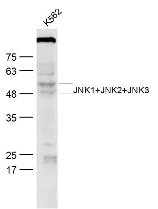
Sample:
K562 (Human) Lysate at 30 ug
Primary: Anti-JNK1+JNK2+JNK3 (bs-2592R) at 1/300 dilution
Secondary: IRDye800CW Goat Anti-Rabbit IgG at 1/20000 dilution
Predicted band size: 42-47 kD
Observed band size:42-52 kD
-
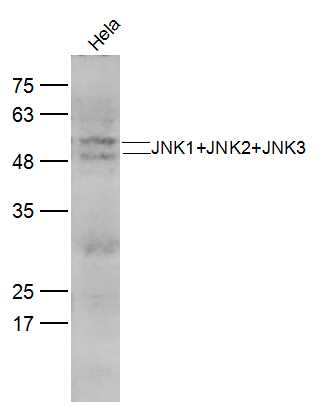
Sample:
Hela(Human) CellLysate at 30 ug
Primary: Anti-JNK1+JNK2+JNK3 (bs-2592R) at 1/300 dilution
Secondary: IRDye800CW Goat Anti-Rabbit IgG at 1/20000 dilution
Predicted band size: 42-47 kD
Observed band size:42-52 kD
-
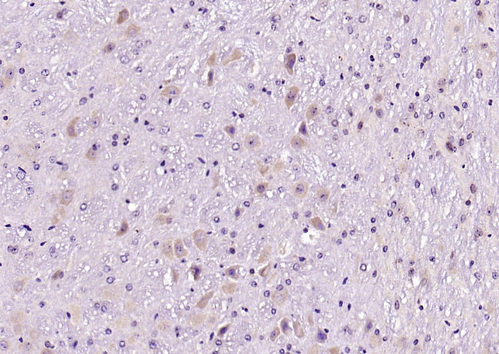
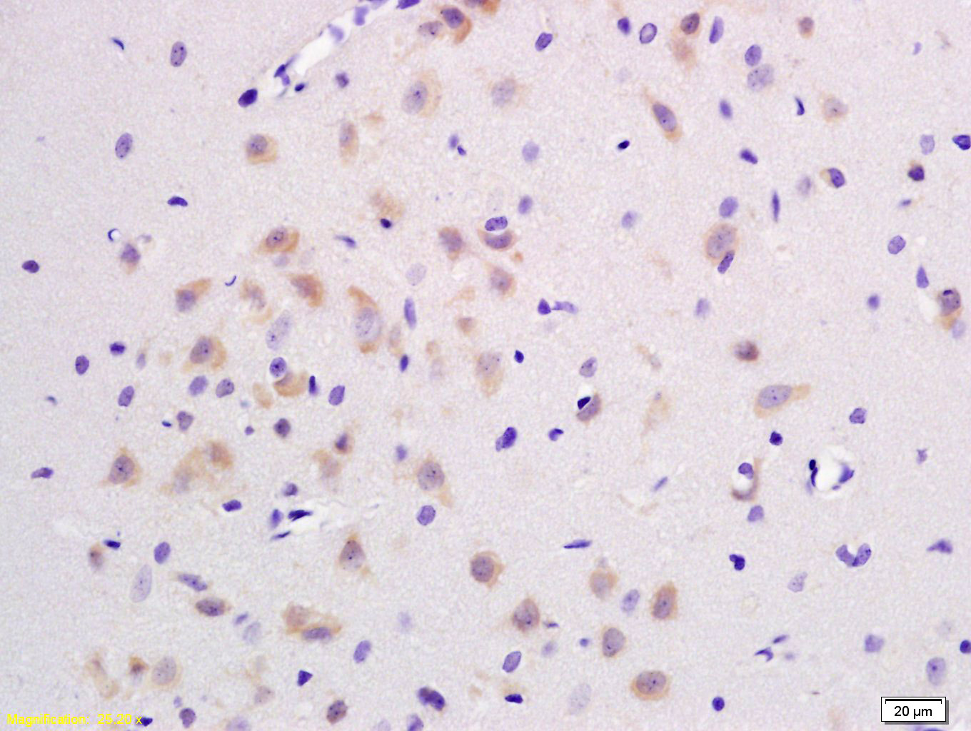
Paraformaldehyde-fixed, paraffin embedded (mouse brain); Antigen retrieval by boiling in sodium citrate buffer (pH6.0) for 15min; Block endogenous peroxidase by 3% hydrogen peroxide for 20 minutes; Blocking buffer (normal goat serum) at 37°C for 30min; Antibody incubation with (JNK1+JNK2+JNK3) Polyclonal Antibody, Unconjugated (bs-2592R) at 1:200 overnight at 4°C, followed by operating according to SP Kit(Rabbit) (sp-0023) instructionsand DAB staining.
-
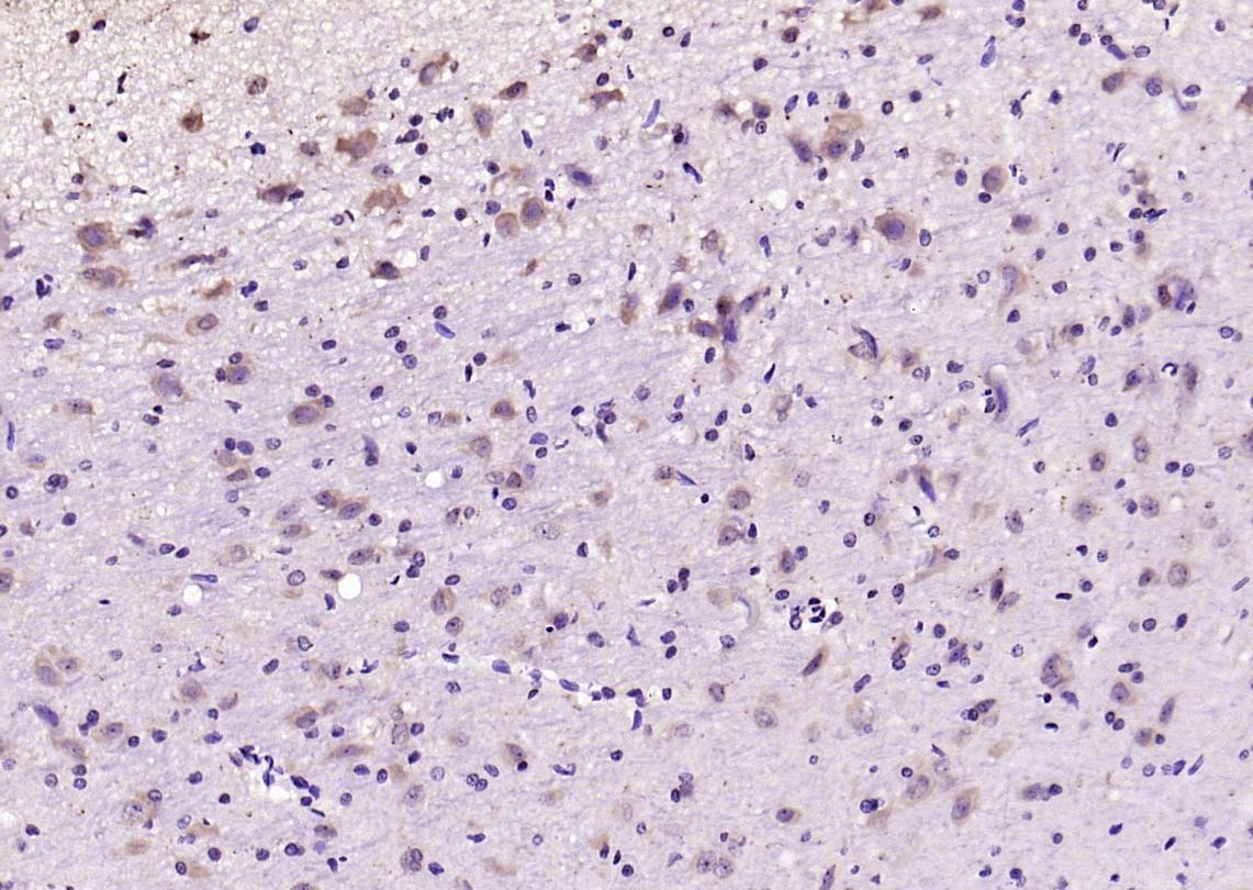
Paraformaldehyde-fixed, paraffin embedded (rat brain); Antigen retrieval by boiling in sodium citrate buffer (pH6.0) for 15min; Block endogenous peroxidase by 3% hydrogen peroxide for 20 minutes; Blocking buffer (normal goat serum) at 37°C for 30min; Antibody incubation with (JNK1+JNK2+JNK3) Polyclonal Antibody, Unconjugated (bs-2592R) at 1:200 overnight at 4°C, followed by operating according to SP Kit(Rabbit) (sp-0023) instructionsand DAB staining.
-
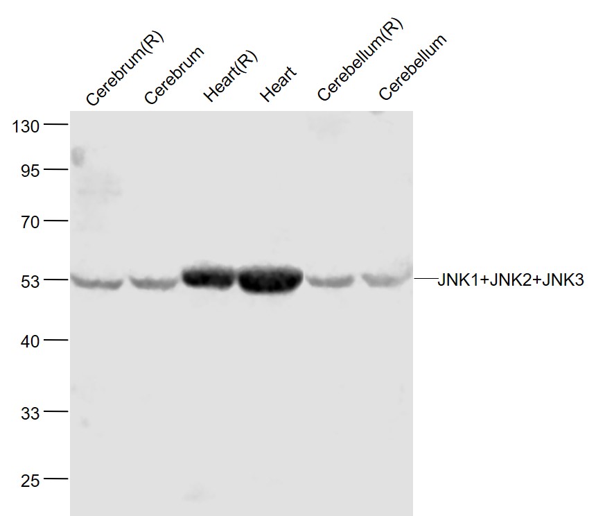
Sample:
Cerebrum(Rat) Lysate at 40 ug
Cerebrum(Mouse) Lysate at 40 ug
Heart(Rat) Lysate at 40 ug
Heart(Mouse) Lysate at 40 ug
Cerebellum(Rat) Lysate at 40 ug
Cerebellum(Mouse) Lysate at 40 ug
Primary: Anti-JNK1+JNK2+JNK3 (bs-2592R) at 1/1000 dilution
Secondary: IRDye800CW Goat Anti-Rabbit IgG at 1/20000 dilution
Predicted band size: 46'54 kD
Observed band size: 54 kD
-
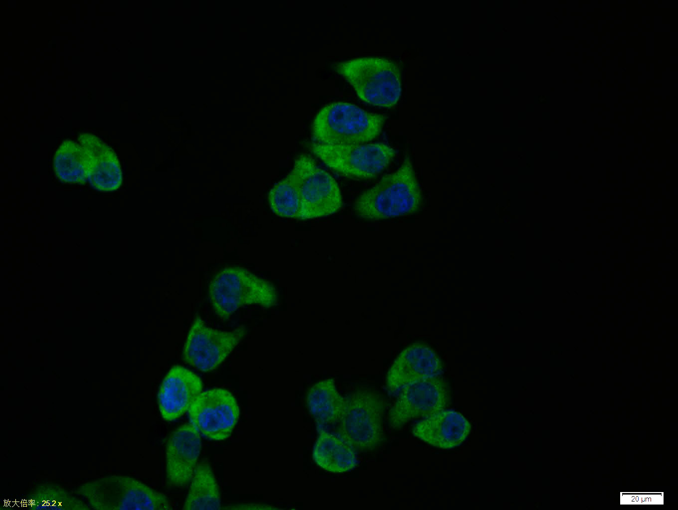
Hela cell; 4% Paraformaldehyde-fixed; Triton X-100 at room temperature for 20 min; Blocking buffer (normal goat serum, C-0005) at 37°C for 20 min; Antibody incubation with (JNK1+JNK2+JNK3) polyclonal Antibody, Unconjugated (bs-2592R) 1:100, 90 minutes at 37°C; followed by a conjugated Goat Anti-Rabbit IgG antibody at 37°C for 90 minutes, DAPI (blue, C02-04002) was used to stain the cell nuclei.
-
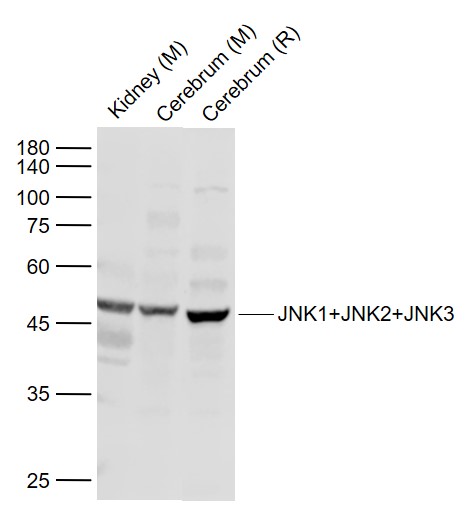
Sample:
Lane 1: Kidney (Mouse) Lysate at 40 ug
Lane 2: Cerebrum (Mouse) Lysate at 40 ug
Lane 3: Cerebrum (Rat) Lysate at 40 ug
Primary: Anti-JNK1+JNK2+JNK3 (bs-2592R) at 1/1000 dilution
Secondary: IRDye800CW Goat Anti-Rabbit IgG at 1/20000 dilution
Predicted band size: 45/54 kD
Observed band size: 48 kD
-
Paraformaldehyde-fixed, paraffin embedded (mouse brain tissue); Antigen retrieval by boiling in sodium citrate buffer (pH6.0) for 15min; Block endogenous peroxidase by 3% hydrogen peroxide for 20 minutes; Blocking buffer (normal goat serum) at 37°C for 30min; Antibody incubation with (JNK1+JNK2+JNK3) Polyclonal Antibody, Unconjugated (bs-2592R) at 1:400 overnight at 4°C, followed by a conjugated secondary (sp-0023) for 20 minutes and DAB staining.
-
Paraformaldehyde-fixed, paraffin embedded (Human kidney); Antigen retrieval by boiling in sodium citrate buffer (pH6.0) for 15min; Block endogenous peroxidase by 3% hydrogen peroxide for 20 minutes; Blocking buffer (normal goat serum) at 37°C for 30min; Antibody incubation with (JNK1+JNK2+JNK3) Polyclonal Antibody, Unconjugated (bs-2592R) at 1:400 overnight at 4°C, followed by operating according to SP Kit(Rabbit) (sp-0023) instructionsand DAB staining.
-
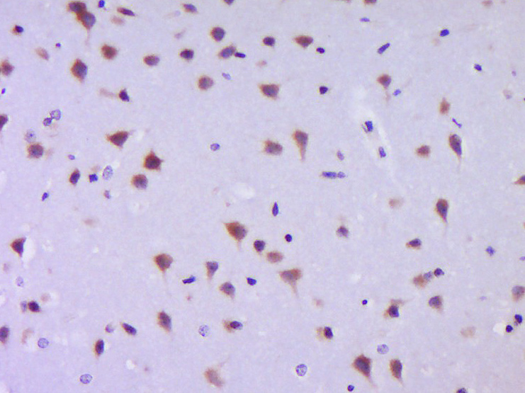
Tissue/cell: rat brain tissue; 4% Paraformaldehyde-fixed and paraffin-embedded;
Antigen retrieval: citrate buffer ( 0.01M, pH 6.0 ), Boiling bathing for 15min; Block endogenous peroxidase by 3% Hydrogen peroxide for 30min; Blocking buffer (normal goat serum,C-0005) at 37℃ for 20 min;
Incubation: Anti-JNK1/2/3 Polyclonal Antibody, Unconjugated(bs-2592R) 1:200, overnight at 4°C, followed by conjugation to the secondary antibody(SP-0023) and DAB(C-0010) staining
-
Sample:
Kidney (Mouse) Lysate at 40 ug
Primary: Anti-JNK1+JNK2+JNK3 (bs-2592R) at 1/300 dilution
Secondary: IRDye800CW Goat Anti-Rabbit IgG at 1/20000 dilution
Predicted band size: 42-47 kD
Observed band size:42-52 kD
-
Blank control (Black line): HUVEC (Black).
Primary Antibody (green line): Rabbit Anti-JNK1+JNK2+JNK3 antibody (bs-2592R)
Dilution: 3μg /10^6 cells;
Isotype Control Antibody (orange line): Rabbit IgG .
Secondary Antibody (white blue line): Goat anti-rabbit IgG-AF647
Dilution: 1μg /test.
Protocol
The cells were fixed with 4% PFA (10min at room temperature)and then permeabilized with 90% ice-cold methanol for 20 min at room temperature. The cells were then incubated in 5%BSA to block non-specific protein-protein interactions for 30 min at room temperature .Cells stained with Primary Antibody for 30 min at room temperature. The secondary antibody used for 40 min at room temperature. Acquisition of 20,000 events was performed.
-
Blank control: Jurkat.
Primary Antibody (green line): Rabbit Anti-JNK1+JNK2+JNK3 antibody (bs-2592R)
Dilution: 1μg /10^6 cells;
Isotype Control Antibody (orange line): Rabbit IgG .
Secondary Antibody : Goat anti-rabbit IgG-AF647
Dilution: 1μg /test.
Protocol
The cells were fixed with 4% PFA (10min at room temperature)and then permeabilized with 90% ice-cold methanol for 20 min at-20℃. The cells were then incubated in 5%BSA to block non-specific protein-protein interactions for 30 min at room temperature .Cells stained with Primary Antibody for 30 min at room temperature. The secondary antibody used for 40 min at room temperature. Acquisition of 20,000 events was performed.
-
Paraformaldehyde-fixed, paraffin embedded (rat cerebellum); Antigen retrieval by boiling in sodium citrate buffer (pH6.0) for 15min; Block endogenous peroxidase by 3% hydrogen peroxide for 20 minutes; Blocking buffer (normal goat serum) at 37°C for 30min; Antibody incubation with (JNK1+JNK2+JNK3) Polyclonal Antibody, Unconjugated (bs-2592R) at 1:200 overnight at 4°C, followed by operating according to SP Kit(Rabbit) (sp-0023) instructionsand DAB staining.
-
Paraformaldehyde-fixed, paraffin embedded (mouse brain); Antigen retrieval by boiling in sodium citrate buffer (pH6.0) for 15min; Block endogenous peroxidase by 3% hydrogen peroxide for 20 minutes; Blocking buffer (normal goat serum) at 37°C for 30min; Antibody incubation with (JNK1+JNK2+JNK3) Polyclonal Antibody, Unconjugated (bs-2592R) at 1:200 overnight at 4°C, followed by operating according to SP Kit(Rabbit) (sp-0023) instructionsand DAB staining.
-
Paraformaldehyde-fixed, paraffin embedded (rat kidney); Antigen retrieval by boiling in sodium citrate buffer (pH6.0) for 15min; Block endogenous peroxidase by 3% hydrogen peroxide for 20 minutes; Blocking buffer (normal goat serum) at 37°C for 30min; Antibody incubation with (JNK1+JNK2+JNK3) Polyclonal Antibody, Unconjugated (bs-2592R) at 1:200 overnight at 4°C, followed by operating according to SP Kit(Rabbit) (sp-0023) instructionsand DAB staining.
-
Paraformaldehyde-fixed, paraffin embedded (mouse cerebellum); Antigen retrieval by boiling in sodium citrate buffer (pH6.0) for 15min; Block endogenous peroxidase by 3% hydrogen peroxide for 20 minutes; Blocking buffer (normal goat serum) at 37°C for 30min; Antibody incubation with (JNK1+JNK2+JNK3) Polyclonal Antibody, Unconjugated (bs-2592R) at 1:200 overnight at 4°C, followed by operating according to SP Kit(Rabbit) (sp-0023) instructionsand DAB staining.
-
Paraformaldehyde-fixed, paraffin embedded (rat brain); Antigen retrieval by boiling in sodium citrate buffer (pH6.0) for 15min; Block endogenous peroxidase by 3% hydrogen peroxide for 20 minutes; Blocking buffer (normal goat serum) at 37°C for 30min; Antibody incubation with (JNK1+JNK2+JNK3) Polyclonal Antibody, Unconjugated (bs-2592R) at 1:200 overnight at 4°C, followed by operating according to SP Kit(Rabbit) (sp-0023) instructionsand DAB staining.
RRID:AB_10883831
产品名称:Rabbit Anti-JNK1+JNK2+JNK3 antibody
别名: JNK1 + JNK2 + JNK3; JNK1/2/3; JNK1+2+3; JNK1 + JNK2 + JNK3; MAPK10; c Jun N terminal kinase 1; c Jun N terminal kinase 2; c Jun N terminal kinase 3; JNK; JNK1; JNK2; JNK2ALPHA; JNK2BETA; JNK3; MAPK8; MAPK9; Mitogen activated protein kinase 10; Mitogen act
中文名称:氨基末端激酶1/2/3抗体
英文名称:Rabbit Anti-JNK1+JNK2+JNK3 antibody
抗体来源: Rabbit
克隆类型:多克隆
细胞定位:细胞核,细胞浆
性 状:Liquid
亚 型:IgG
纯化方法:affinity purified by Protein A
保存条件:Shipped at 4℃. Store at -20 °C for one year. Avoid repeated freeze/thaw cycles.
免 疫 原:KLH conjugated synthetic peptide derived from human JNK1/2/3
抗原表位:151-250/384
SWISS:Q61831
Gene ID :5599
Human Gene ID:5599
JNK1(MAPK8) is a member of the MAP kinase family. JNK1 is activated by threonine and tyrosine phosphorylation by either of two dual specificity kinases, MAP2K4 and MAP2K7.
JNK2 (p54a, SAPK1a), along with JNK1 and JNK3, is thought to play an important role in nuclear signal transduction through its environmental stress activation and subsequent phosphorylation of the nuclear transcription factor p53.
JNK3 is a neuron-specific form of c-Jun N-terminal kinases. Through its phosphorylation and nuclear localization, this kinase plays regulatory roles in the signaling pathways of neuronal apoptosis.
The JNK pathway is critically involved in diabetes and levels are abnormally elevated in obesity.
Important Note:This product as supplied is intended for research use only, not for use in human, therapeutic or diagnostic applications.
 400-901-9800


 说明书
说明书 联系我们
联系我们 打印此页面
打印此页面 收藏
收藏