 400-901-9800


|
|
多克隆 |
SKU:bs-1712R
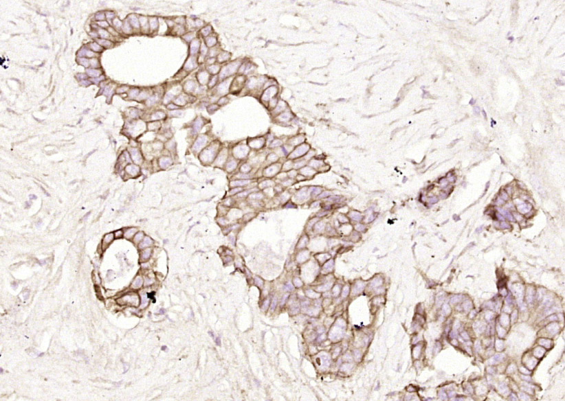
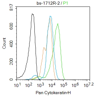
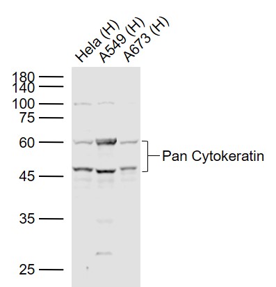
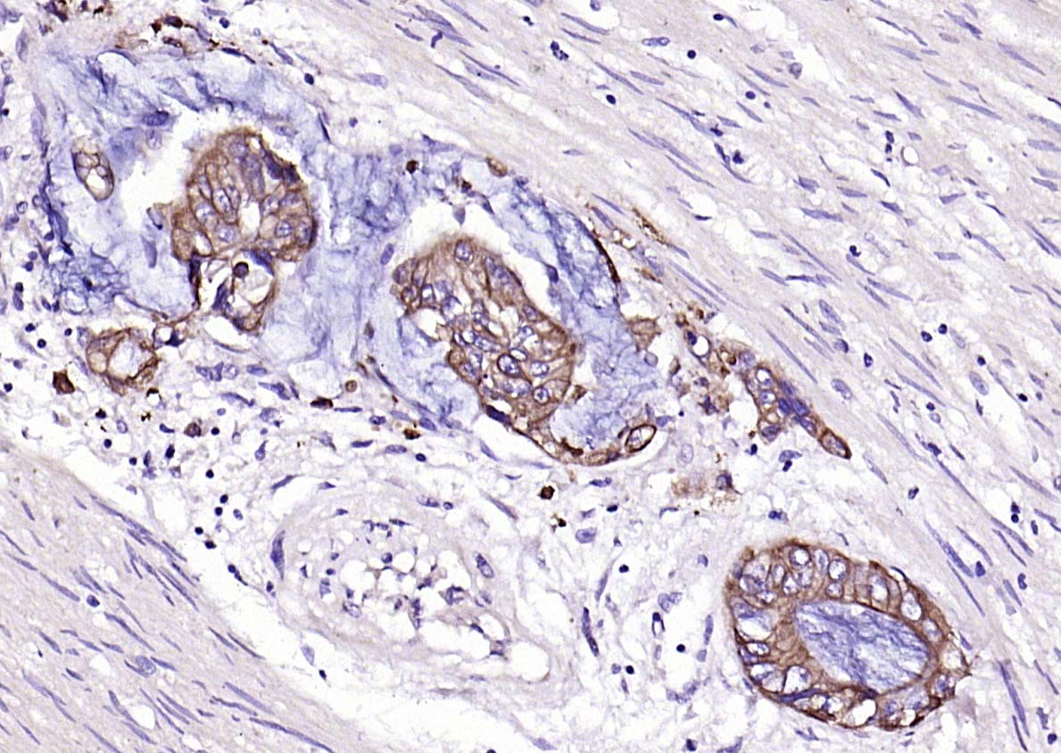
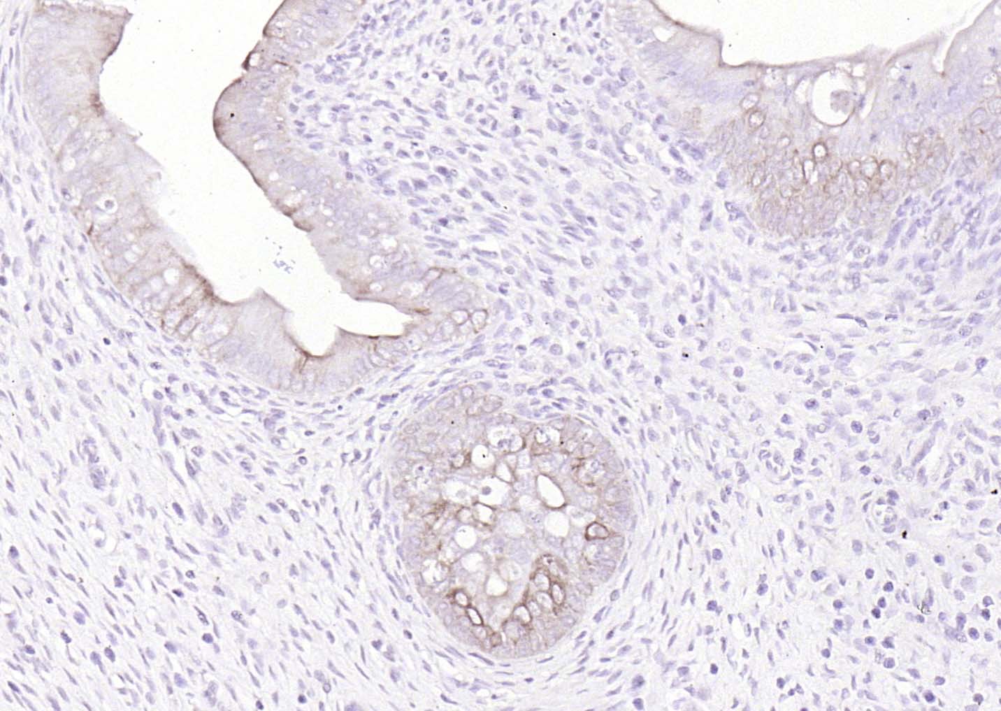
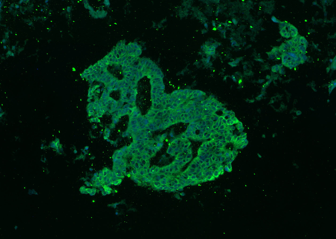
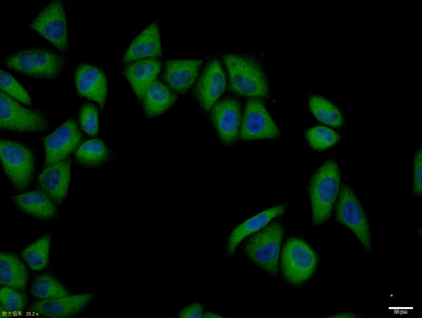
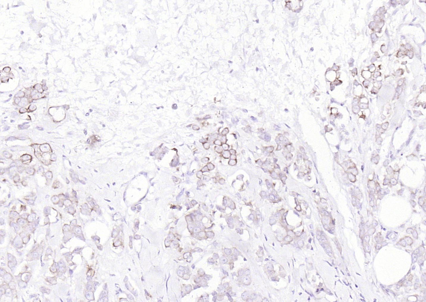
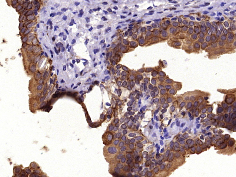
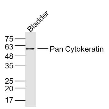
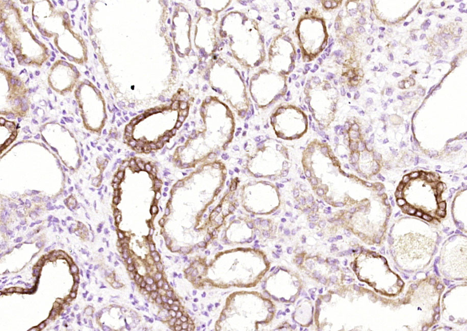
| Rabbit Anti-Pan Cytokeratin antibody | |
| 反应物种(预测) | Chicken,Dog,Pig,Horse,Rabbit |
| 产品应用(已验证) | WB,IHC,ICC,IF,FCM |
| 产品应用(可尝试) | ELISA |
| 推荐稀释比例 | WB=1:500-2000,Elisa=1:5000-10000,IHC-P=1:100-500,IHC-F=1:100-500,Flow Cyt=1μg /test,IF=1:100-500,ICC=1:100, |
| 研究领域 | 肿瘤,细胞生物,免疫学 |
| 标签 | Array |


产品信息
免疫原信息
产品介绍
 说明书
说明书 联系我们
联系我们 打印此页面
打印此页面 收藏
收藏