| Rabbit Anti-phospho-Bax (Ser184) antibody |
| 反应物种(预测) |
Dog,Pig,Cow,Sheep |
| 产品应用(已验证) |
WB,IHC,FCM |
| 产品应用(可尝试) |
ELISA |
| 推荐稀释比例 |
WB=1:500-2000,Elisa=1:5000-10000,IHC-P=1:100-500,Flow Cyt=3μg /test, |
| 研究领域 |
肿瘤,细胞生物,神经生物学,信号转导,细胞凋亡 |
| 标签 |
Array |
-
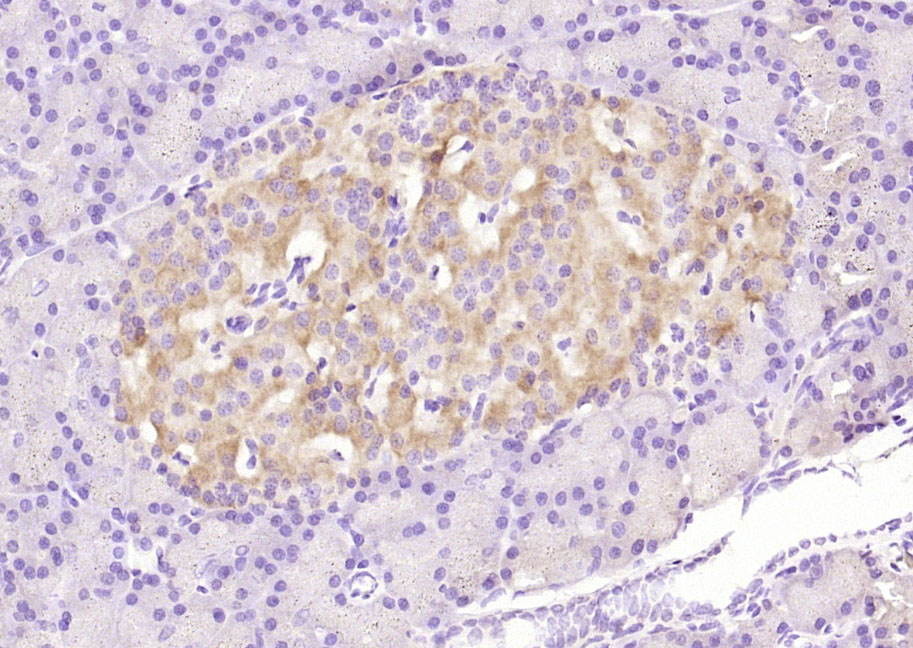
Paraformaldehyde-fixed, paraffin embedded (rat pancreas); Antigen retrieval by boiling in sodium citrate buffer (pH6.0) for 15min; Block endogenous peroxidase by 3% hydrogen peroxide for 20 minutes; Blocking buffer (normal goat serum) at 37°C for 30min; Antibody incubation with (phospho-Bax (Ser184)) Polyclonal Antibody, Unconjugated (bs-3010R) at 1:200 overnight at 4°C, followed by operating according to SP Kit(Rabbit) (sp-0023) instructionsand DAB staining.
-
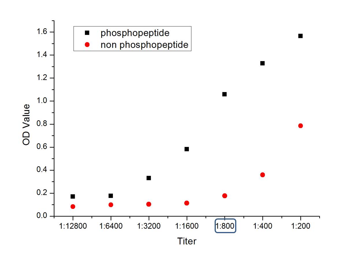
phosphopeptide
non phosphopeptide
-
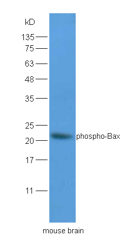
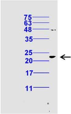
Sample:Brain(Mouse) lysate at 30ug;
Primary: Anti-phospho-Bax (Ser184) (bs-3010R) at 1:200 dilution;
Secondary: HRP conjugated Goat Anti-Rabbit IgG(bs-0295G-HRP) at 1: 5000 dilution;
Predicted band size : 21kD
Observed band size : 21kD
-
Tissue/cell: rat brain tissue;4% Paraformaldehyde-fixed and paraffin-embedded;
Antigen retrieval: citrate buffer ( 0.01M, pH 6.0 ), Boiling bathing for 15min; Blocking buffer (normal goat serum,C-0005) at 37℃ for 20 min;
Incubation: Anti-phospho-Bax(Ser184) Polyclonal Antibody, Unconjugated(bs-3010R) 1:200, overnight at 4°C; The secondary antibody was Goat Anti-Rabbit IgG, Cy3 conjugated (bs-0295G-Cy3)used at 1:200 dilution for 40 minutes at 37°C. DAPI(5ug/ml,blue,C-0033) was used to stain the cell nuclei
-
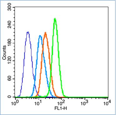
Blank control (blue line): HL60 (fixed with 2% paraformaldehyde (10 min) and then permeabilized with 0.1% PBS-Tween for 20 min at room temperature).
Primary Antibody (green line): Rabbit Anti-phospho-Bax (Ser184) antibody (bs-3010R),Dilution: 3μg /10^6 cells;
Isotype Control Antibody (orange line): Rabbit IgG .
Secondary Antibody (white blue line): Goat anti-rabbit IgG-FITC,Dilution: 1μg /test.
-
Sample: Testis (Mouse) Tissue Lysate at 30 ug
Primary: Anti-phospho-Bax (Ser184) (bs-3010R)at 1/300 dilution
Secondary: IRDye800CW Goat Anti-Rabbit IgG at 1/20000 dilution
Predicted band size: 21 kD
Observed band size: 22 kD
RRID:AB_10856175
产品名称:Rabbit Anti-phospho-Bax (Ser184) antibody
别名: Bax (phospho S184); Bax (phospho Ser184); p-Bax (S184); p-Bax (Ser184); apoptosis regulator BAX; Apoptosis regulator BAX cytoplasmic isoform beta; Apoptosis regulator BAX membrane isoform alpha; Bax isoform psi; BAX protein cytoplasmic isoform delta; Bax
中文名称:磷酸化Bax抗体
英文名称:Rabbit Anti-phospho-Bax (Ser184) antibody
抗体来源: Rabbit
克隆类型:多克隆
细胞定位:细胞浆,细胞膜
性 状:Liquid
亚 型:IgG
纯化方法:affinity purified by Protein A
保存条件:Shipped at 4℃. Store at -20 °C for one year. Avoid repeated freeze/thaw cycles.
免 疫 原:KLH conjugated Synthesised phosphopeptide derived from human Bax around the phosphorylation site of Ser184
抗原表位:TA(p-S)LT
SWISS:Q07812
Gene ID :581
Human Gene ID:581
The protein encoded by this gene belongs to the BCL2 protein family. BCL2 family members form hetero- or homodimers and act as anti- or pro-apoptotic regulators that are involved in a wide variety of cellular activities. This protein forms a heterodimer with BCL2, and functions as an apoptotic activator. This protein is reported to interact with, and increase the opening of, the mitochondrial voltage-dependent anion channel (VDAC), which leads to the loss in membrane potential and the release of cytochrome c. The expression of this gene is regulated by the tumor suppressor P53 and has been shown to be involved in P53-mediated apoptosis. Multiple alternatively spliced transcript variants, which encode different isoforms, have been reported for this gene. [provided by RefSeq, Jul 2008].
Function:Accelerates programmed cell death by binding to, and antagonizing the apoptosis repressor BCL2 or its adenovirus homolog E1B 19k protein. Under stress conditions, undergoes a conformation change that causes translocation to the mitochondrion membrane, lea
Subunit:Homodimer. Forms higher oligomers under stress conditions. Interacts with BCL2L11. Interaction with BCL2L11 promotes BAX oligomerization and association with mitochondrial membranes, with subsequent release of cytochrome c. Forms heterodimers with BCL2, E
Subcellular Location:Isoform Alpha: Mitochondrion membrane; Single-pass membrane protein. Cytoplasm. Note=Colocalizes with 14-3-3 proteins in the cytoplasm. Under stress conditions, undergoes a conformation change that causes release from JNK-phosphorylated 14-3-3 proteins an
Tissue Specificity:Expressed in a wide variety of tissues. Isoform Psi is found in glial tumors. Isoform Alpha is expressed in spleen, breast, ovary, testis, colon and brain, and at low levels in skin and lung. Isoform Sigma is expressed in spleen, breast, ovary, testis, lu
Similarity:Belongs to the Bcl-2 family.
Important Note:This product as supplied is intended for research use only, not for use in human, therapeutic or diagnostic applications.
 400-901-9800


 说明书
说明书 联系我们
联系我们 打印此页面
打印此页面 收藏
收藏