欢迎访问博奥森抗体公司官方网站!网站测试中
首页 > 产品 > 一抗 > 产品信息
Rabbit Anti-FAK antibody
多克隆  |   SKU:bs-1340R

说明书

联系我们

打印此页面

收藏

货号:bs-3351R
¥1280
订购号:bs-1340R
¥1098.00-2900.00
货期:现货
Rabbit Anti-FAK antibody
产品应用(已验证)

WB,IHC,ICC,IF,FCM

产品应用(可尝试)

ELISA

推荐稀释比例

WB=1:500-2000,Elisa=1:5000-10000,IHC-P=1:100-500,IHC-F=1:100-500,Flow Cyt=1μg/Test,IF=1:100-500,ICC=1:100,

研究领域

肿瘤,细胞生物,免疫学,激酶和磷酸酶

标签

Array

  • Sample:
    MCF-7(Human) Cell Lysate at 30 ug
    Primary: Anti-FAK (bs-1340R) at 1/300 dilution
    Secondary: IRDye800CW Goat Anti-Rabbit IgG at 1/20000 dilution
    Predicted band size: 116 kD
    Observed band size: 116 kD
  • HUVEC cell; 4% Paraformaldehyde-fixed; Triton X-100 at room temperature for 20 min; Blocking buffer (normal goat serum, C-0005) at 37°C for 20 min; Antibody incubation with (FAK) polyclonal Antibody, Unconjugated (bs-1340R) 1:100, 90 minutes at 37°C; followed by a conjugated Goat Anti-Rabbit IgG antibody at 37°C for 90 minutes, DAPI (blue, C02-04002) was used to stain the cell nuclei.
  • Tissue/cell: mouse lung tissue;4% Paraformaldehyde-fixed and paraffin-embedded;
    Antigen retrieval: citrate buffer ( 0.01M, pH 6.0 ), Boiling bathing for 15min; Blocking buffer (normal goat serum,C-0005) at 37℃ for 20 min;
    Incubation: Anti-FAK/PTK2 Polyclonal Antibody, Unconjugated(bs-1340R) 1:200, overnight at 4°C; The secondary antibody was Goat Anti-Rabbit IgG, Cy3 conjugated(bs-0295G-Cy3)used at 1:200 dilution for 40 minutes at 37°C. DAPI(5ug/ml,blue,C-0033) was used to stain the cell nuclei
  • Sample:Thymus (Mouse) Lysate at 40 ug
    Primary: Anti-FAK(bs-1340R)at 1/300 dilution
    Secondary: IRDye800CW Goat Anti-Rabbit IgG at 1/20000 dilution
    Predicted band size: 116kD
    Observed band size: 116kD
  • Paraformaldehyde-fixed, paraffin embedded (Mouse brain); Antigen retrieval by boiling in sodium citrate buffer (pH6.0) for 15min; Block endogenous peroxidase by 3% hydrogen peroxide for 20 minutes; Blocking buffer (normal goat serum) at 37°C for 30min; Antibody incubation with (FAK) Polyclonal Antibody, Unconjugated (bs-1340R) at 1:400 overnight at 4°C, followed by operating according to SP Kit(Rabbit) (sp-0023) instructionsand DAB staining.
  • Blank control (blue line): Hep G2 (blue).
    Primary Antibody (green line): Rabbit Anti-FAK antibody (bs-1340R)
    Dilution: 1μg /10^6 cells;
    Isotype Control Antibody (orange line): Rabbit IgG .
    Secondary Antibody (white blue line): Goat anti-rabbit IgG-PE
    Dilution: 1μg /test.
    Protocol
    The cells were fixed with 70% ethanol (Overmight at 4℃) and then permeabilized with 90% ice-cold methanol for 30 min at -20℃.Cells stained with Primary Antibody for 30 min at room temperature. The cells were then incubated in 1 X PBS/2%BSA/10% goat serum to block non-specific protein-protein interactions followed by the antibody for 15 min at room temperature. The secondary antibody used for 40 min at room temperature. Acquisition of 20,000 events was performed.
  • Sample:
    Lane 1: Cerebrum (Mouse) Lysate at 40 ug
    Lane 2: NIH/3T3 (Mouse) Cell Lysate at 30 ug
    Lane 3: Cerebrum (Rat) Lysate at 40 ug
    Lane 4: 293T (Human) Cell Lysate at 30 ug
    Lane 5: A431 (Human) Cell Lysate at 30 ug
    Lane 6: Huvec (Human) Cell Lysate at 30 ug
    Lane 7: A549 (Human) Cell Lysate at 30 ug
    Primary: Anti-FAK (bs-1340R) at 1/1000 dilution
    Secondary: IRDye800CW Goat Anti-Rabbit IgG at 1/20000 dilution
    Predicted band size: 125 kD
    Observed band size: 120 kD

产品信息

RRID:AB_10857573
产品名称:Rabbit Anti-FAK antibody
别名: FADK 1; FADK; FAK 1; FAK related non kinase polypeptide; FAK1; Focal adhesion kinase 1; FRNK; pp125FAK; Protein tyrosine kinase 2; Protein Tyrosine Kinase Cytoplasmic; PTK 2; FAK1_HUMAN; Focal adhesion kinase-related nonkinase; Protein phosphatase 1 regul
中文名称:粘着斑激酶抗体
英文名称:Rabbit Anti-FAK antibody
抗体来源: Rabbit
克隆类型:多克隆
细胞定位:细胞核,细胞浆,细胞膜
性 状:Liquid
亚 型:IgG
纯化方法:affinity purified by Protein A
保存条件:Shipped at 4℃. Store at -20 °C for one year. Avoid repeated freeze/thaw cycles.

免疫原信息

免 疫 原:KLH conjugated synthetic peptide derived from human FAK
抗原表位:901-1052/1052
SWISS:Q05397
Gene ID :5747
Human Gene ID:5747

产品介绍

This gene encodes a cytoplasmic protein tyrosine kinase which is found concentrated in the focal adhesions that form between cells growing in the presence of extracellular matrix constituents. The encoded protein is a member of the FAK subfamily of protein tyrosine kinases but lacks significant sequence similarity to kinases from other subfamilies. Activation of this gene may be an important early step in cell growth and intracellular signal transduction pathways triggered in response to certain neural peptides or to cell interactions with the extracellular matrix. Several transcript variants encoding different isoforms have been found for this gene, but the full-length natures of only three of them have been determined. [provided by RefSeq, Dec 2010]
Function:Non-receptor protein-tyrosine kinase that plays an essential role in regulating cell migration, adhesion, spreading, reorganization of the actin cytoskeleton, formation and disassembly of focal adhesions and cell protrusions, cell cycle progression, cell
Subunit:Interacts (via first Pro-rich region) with CAS family members (via SH3 domain), including BCAR1, BCAR3, CASS4 and NEDD9. Interacts with GIT1. Interacts with SORBS1. Interacts with RGNEF. Interacts with SHB. Interacts with PXN and TLN1. Interacts with STAT
Subcellular Location:Cell junction, focal adhesion. Cell membrane; Peripheral membrane protein; Cytoplasmic side. Cytoplasm, cell cortex. Cytoplasm, cytoskeleton. Cytoplasm, cytoskeleton, centrosome. Nucleus. Note=Constituent of focal adhesions. Detected at microtubules.
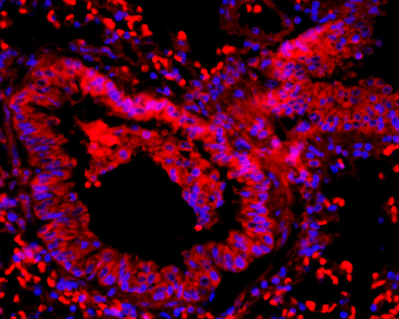
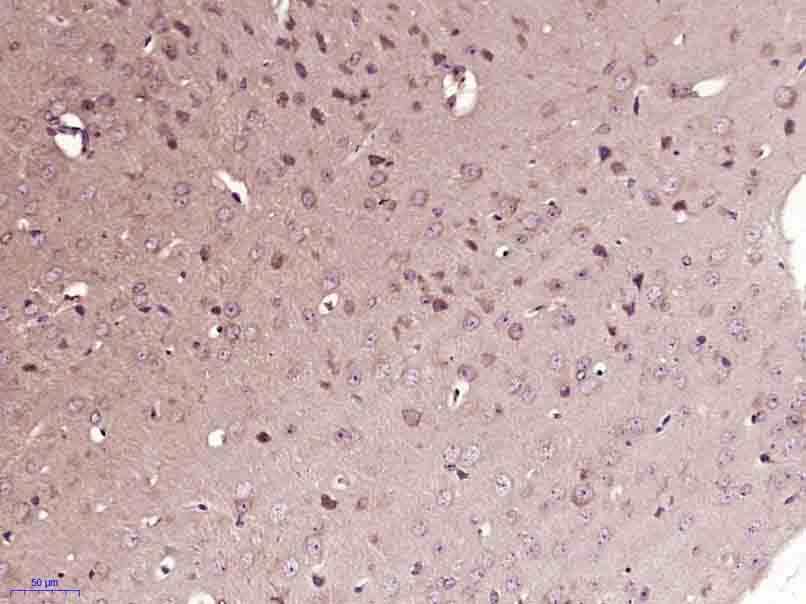
Tissue Specificity:Detected in B and T-lymphocytes. Isoform 1 and isoform 6 are detected in lung fibroblasts (at protein level). Ubiquitous.
Post-translational modifications:Phosphorylated on tyrosine residues upon activation, e.g. upon integrin signaling. Tyr-397 is the major autophosphorylation site, but other kinases can also phosphorylate this residue. Phosphorylation at Tyr-397 promotes interaction with SRC and SRC famil
DISEASE:Note=Aberrant PTK2/FAK1 expression may play a role in cancer cell proliferation, migration and invasion, in tumor formation and metastasis. PTK2/FAK1 overexpression is seen in many types of cancer.
Similarity:Belongs to the protein kinase superfamily. Tyr protein kinase family.
FAK subfamily.
Contains 1 FERM domain.

Important Note:This product as supplied is intended for research use only, not for use in human, therapeutic or diagnostic applications.

产品订购:sales@bioss.com.cn
邮政编码:101102
综合邮箱:bioss@bioss.com.cn
联系地址:北京市通州区马驹桥镇景盛南四街联东U谷西区2号院67号楼
版权所有 2019-2023 www.bioss.com.cn 北京博奥森生物技术有限公司 京ICP备05066980号-8 京公网安备110107000727号