| Rabbit Anti-phospho-GSK-3 Beta (Ser9) antibody |
| 产品应用(已验证) |
WB,IHC,ICC,FCM |
| 产品应用(可尝试) |
IF,ELISA |
| 推荐稀释比例 |
WB=1:500-2000,Elisa=1:5000-10000,IHC-P=1:100-500,IHC-F=1:100-500,Flow Cyt=1ug/Test,IF=1:100-500,ICC=1:100, |
| 研究领域 |
细胞生物,神经生物学,信号转导,细胞凋亡,激酶和磷酸酶 |
| 标签 |
Array |
-
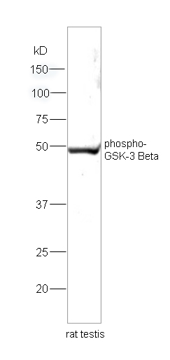
Sample: Testis (Rat) Lysate at 30 ug
Primary: Anti-phospho-GSK-3 Beta(Ser9) (bs-2066R) at 1:200 dilution;
Secondary: HRP conjugated Goat Anti-Rabbit IgG(bs-0295G-HRP) at 1: 5000 dilution;
Predicted band size : 47kD
Observed band size : 49kD
-
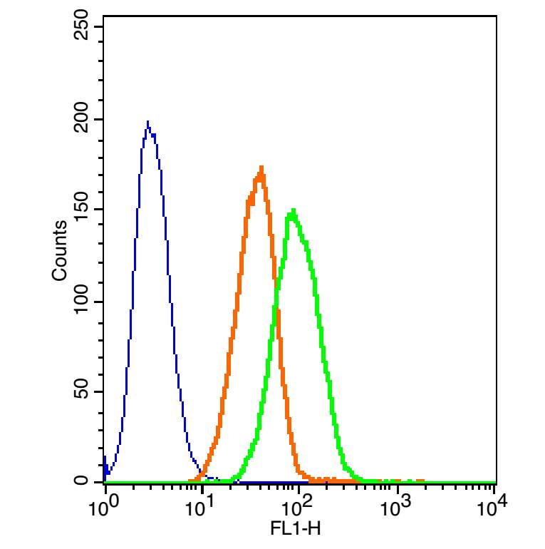
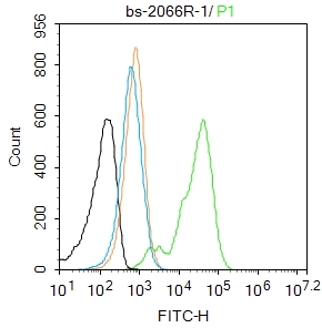
The blue histogram is unstained cells(A549 cells).
The Orange histogram is cells stained with Rabbit IgG/FITC (bs-0295P-FITC).
The green histogram is cells stained with Rabbit Anti-phospho-GSK-3 Beta(Ser9)/FITC Conjugated antibody (bs-2066R-FITC).
Isotype control: Cell lines treated with Rabbit IgG/FITC(bs-0295P-FITC) instead of the primary antibody to confirm that primary antibody binding is specific. Concentration: 5μL in 100 μL 1 X PBS containing 0.5% BSA.
-
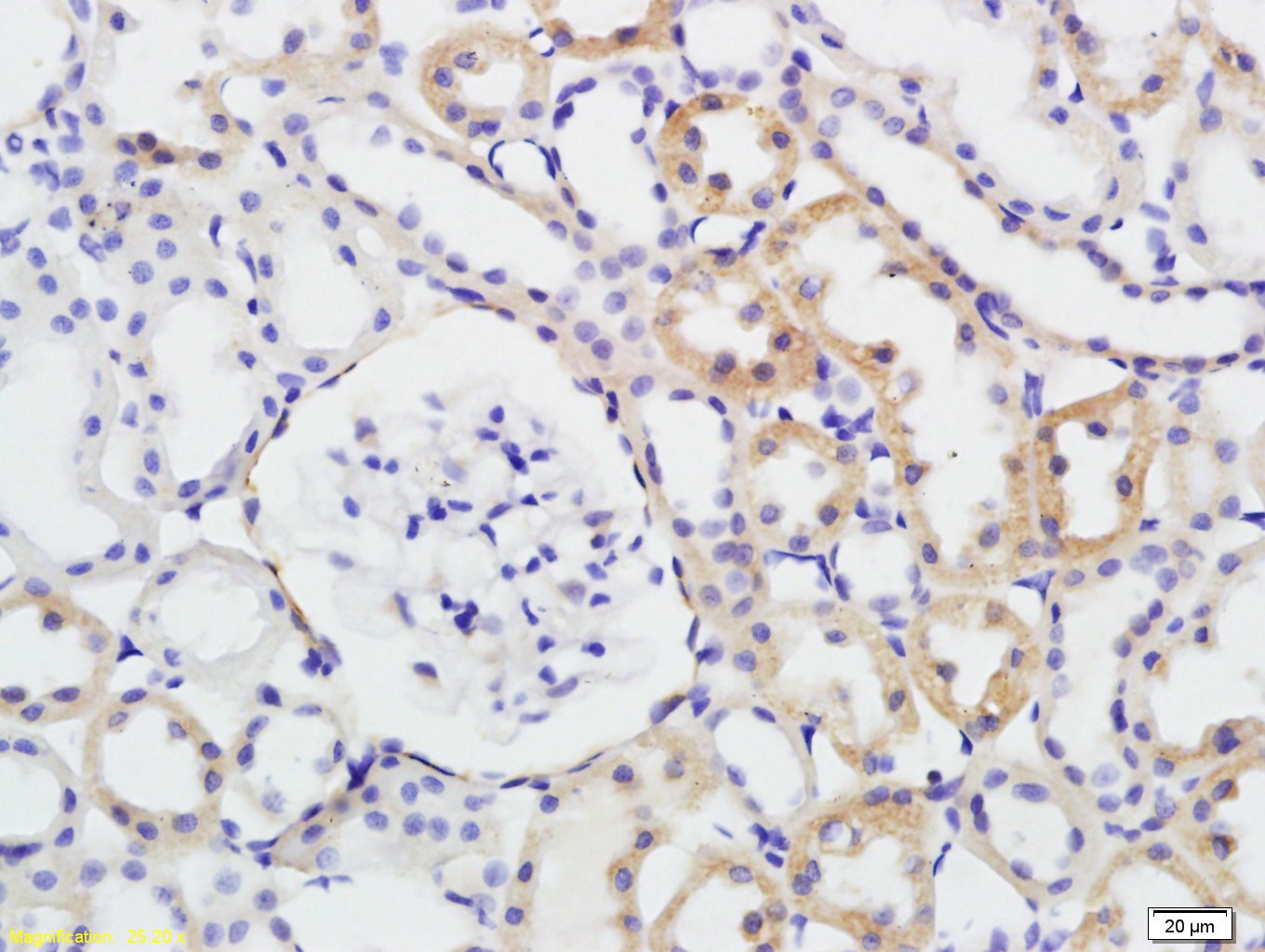
Tissue/cell: rat kidney tissue; 4% Paraformaldehyde-fixed and paraffin-embedded;
Antigen retrieval: citrate buffer ( 0.01M, pH 6.0 ), Boiling bathing for 15min; Block endogenous peroxidase by 3% Hydrogen peroxide for 30min; Blocking buffer (normal goat serum,C-0005) at 37℃ for 20 min;
Incubation: Anti-phospho-GSK-3 Beta(Ser9) Polyclonal Antibody, Unconjugated(bs-2066R) 1:200, overnight at 4°C, followed by conjugation to the secondary antibody(SP-0023) and DAB(C-0010) staining
-
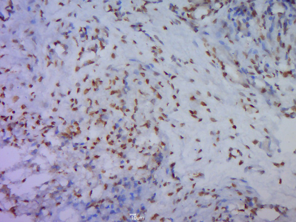
Paraformaldehyde-fixed, paraffin embedded (Mouse placenta); Antigen retrieval by boiling in sodium citrate buffer (pH6.0) for 15min; Block endogenous peroxidase by 3% hydrogen peroxide for 20 minutes; Blocking buffer (normal goat serum) at 37°C for 30min; Antibody incubation with (Beta(Ser9)) Polyclonal Antibody, Unconjugated (bs-2066R p-GSK-3) at 1:500 overnight at 4°C, followed by a conjugated secondary (sp-0023) for 20 minutes and DAB staining.
-
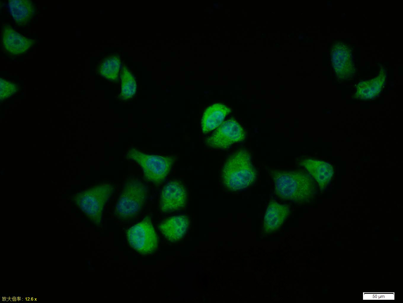
Tissue/cell:Hela cell; 4% Paraformaldehyde-fixed; Triton X-100 at room temperature for 20 min; Blocking buffer (normal goat serum, C-0005) at 37°C for 20 min; Antibody incubation with (phospho-GSK-3 Beta (Ser9)) polyclonal Antibody, Unconjugated (bs-2066R) 1:100, 90 minutes at 37°C; followed by a FITC conjugated Goat Anti-Rabbit IgG antibody at 37°C for 90 minutes, DAPI (blue, C02-04002) was used to stain the cell nuclei.
-
Blank control: A431.
Primary Antibody (green line): Rabbit Anti-phospho-GSK-3 Beta (Ser9) antibody (bs-2066R)
Dilution: 1μg /10^6 cells;
Isotype Control Antibody (orange line): Rabbit IgG .
Secondary Antibody : Goat anti-rabbit IgG-AF488
Dilution: 1μg /test.
Protocol
The cells were fixed with 4% PFA (10min at room temperature)and then permeabilized with 90% ice-cold methanol for 20 min at-20℃. The cells were then incubated in 5%BSA to block non-specific protein-protein interactions for 30 min at room temperature .Cells stained with Primary Antibody for 30 min at room temperature. The secondary antibody used for 40 min at room temperature. Acquisition of 20,000 events was performed.
RRID:AB_10857487
产品名称:Rabbit Anti-phospho-GSK-3 Beta (Ser9) antibody
别名: GSK3 beta (phospho S9); p-GSK3 beta (phospho S9); GSK3B(Phospho-Ser9); GSK3B(Phospho-S9); p-GSK-3 Beta(Ser9); p-GSK-3 beta(S9); Glycogen synthase kinase 3 beta; GSK 3 beta; GSK 3B; GSK3B; GSK3B protein; GSK3beta isoform; GSK3 beta; Glycogen synthase kinas
中文名称:磷酸化糖原合酶激酶-3β抗体
英文名称:Rabbit Anti-phospho-GSK-3 Beta (Ser9) antibody
抗体来源: Rabbit
克隆类型:多克隆
细胞定位:细胞核,细胞浆,细胞膜
性 状:Liquid
亚 型:IgG
纯化方法:affinity purified by Protein A
保存条件:Shipped at 4℃. Store at -20 °C for one year. Avoid repeated freeze/thaw cycles.
免 疫 原:KLH conjugated Synthesised phosphopeptide derived from human GSK-3 Beta around the phosphorylation site of Ser9
抗原表位:TT(p-S)FA
SWISS:P49841
Gene ID :2932
Human Gene ID:2932
The protein encoded by this gene is a serine-threonine kinase, belonging to the glycogen synthase kinase subfamily. It is involved in energy metabolism, neuronal cell development, and body pattern formation. Polymorphisms in this gene have been implicated in modifying risk of Parkinson disease, and studies in mice show that overexpression of this gene may be relevant to the pathogenesis of Alzheimer disease. Alternatively spliced transcript variants encoding different isoforms have been found for this gene.[provided by RefSeq, Sep 2009]
Function:Constitutively active protein kinase that acts as a negative regulator in the hormonal control of glucose homeostasis, Wnt signaling and regulation of transcription factors and microtubules, by phosphorylating and inactivating glycogen synthase (GYS1 or G
Subunit:Monomer. Interacts with ARRB2 and DISC1. Interacts with CABYR, MMP2, MUC1, NIN and PRUNE Interacts with AXIN1; the interaction mediates hyperphosphorylation of CTNNB1 leading to its ubiquitination and destruction. Interacts with and phosphorylates SNAI1.
Subcellular Location:Cytoplasm. Nucleus. Cell membrane. Note=The phosphorylated form shows localization to cytoplasm and cell membrane. The MEMO1-RHOA-DIAPH1 signaling pathway controls localization of the phosophorylated form to the cell membrane.
Tissue Specificity:Expressed in testis, thymus, prostate and ovary and weakly expressed in lung, brain and kidney.
Post-translational modifications:Phosphorylated by AKT1 and ILK1. Upon insulin-mediated signaling, the activated PKB/AKT1 protein kinase phosphorylates and desactivates GSK3B, resulting in the dephosphorylation and activation of GYS1. Activated by phosphorylation at Tyr-216.
Similarity:Belongs to the protein kinase superfamily. CMGC Ser/Thr protein kinase family. GSK-3 subfamily.
Contains 1 protein kinase domain.
Important Note:This product as supplied is intended for research use only, not for use in human, therapeutic or diagnostic applications.
 400-901-9800


 说明书
说明书 联系我们
联系我们 打印此页面
打印此页面 收藏
收藏