| Rabbit Anti-CD63 antibody |
| 产品应用(已验证) |
WB,IHC,FCM |
| 产品应用(可尝试) |
ICC,IF,ELISA |
| 推荐稀释比例 |
WB=1:500-2000,Elisa=1:5000-10000,IHC-P=1:100-500,IHC-F=1:100-500,Flow Cyt=1μg/Test,IF=1:100-500,ICC=1:100-500, |
| 研究领域 |
肿瘤,心血管,免疫学,细胞类型标志物(Tumor biomarkers; Platelets markers.), |
| 标签 |
Array |
-
cell: A549 cells
Incubation: Avoid light, at 4°C for 60 minutes.
Blank control A549 cells(red line),5X10^5/ml, at 4°C for 60 minutes.
primary antibody: abbit Anti-CD63 antibody (bs-1523R-PE,Blue line) ,1:50, at 4°C for 60 minutes.
-
Blank control: Hep G2 cells(blue).
Primary Antibody:Rabbit Anti- CD23 antibody(bs-1523R), Dilution: 1μg in 100 μL 1X PBS containing 0.5% BSA;
Isotype Control Antibody: Rabbit IgG(orange) ,used under the same conditions );
Secondary Antibody: Goat anti-rabbit IgG-PE(white blue), Dilution: 1:200 in 1 X PBS containing 0.5% BSA.
Protocol
The cells were fixed with 2% paraformaldehyde (10 min) , then permeabilized with 90% ice-cold methanol for 30 min on ice. Primary antibody (bs-1523R,1μg /1x10^6 cells) were incubated for 30 min on the ice, followed by 1 X PBS containing 0.5% BSA + 1 0% goat serum (15 min) to block non-specific protein-protein interactions. Then the Goat Anti-rabbit IgG/PE antibody was added into the blocking buffer mentioned above to react with the primary antibody at 1/200 dilution for 30 min on ice. Acquisition of 20,000 events was performed.
-
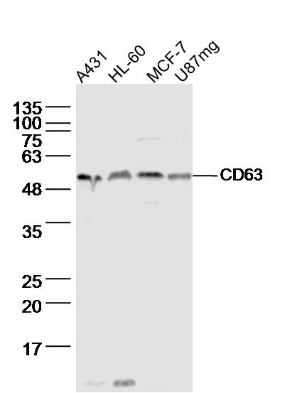
Sample:
A431 cell(human) Lysate at 40 ug
HL-60 cell(human) Lysate at 40 ug
MCF-7 cell(human) Lysate at 40 ug
U87mg cell(human)Lysate at 40 ug
Primary: Anti- CD63 (bs-1523R)at 1/300 dilution
Secondary: IRDye800CW Goat Anti-Rabbit IgG at 1/20000 dilution
Predicted band size: 26kD
Observed band size: 53 kD
-
Tissue/cell: human lung carcinoma; 4% Paraformaldehyde-fixed and paraffin-embedded;
Antigen retrieval: citrate buffer ( 0.01M, pH 6.0 ), Boiling bathing for 15min; Block endogenous peroxidase by 3% Hydrogen peroxide for 30min; Blocking buffer (normal goat serum,C-0005) at 37℃ for 20 min;
Incubation: Anti-CD63 Polyclonal Antibody, Unconjugated(bs-1523R) 1:200, overnight at 4°C, followed by conjugation to the secondary antibody(SP-0023) and DAB(C-0010) staining
RRID:AB_10857113
产品名称:Rabbit Anti-CD63 antibody
别名: Lysosomal associated membrane protein 3; CD63 antigen; CD63 antigen melanoma 1 antigen; CD63 molecule; granulophysin; LAMP 3; LAMP3; lysosome associated membrane glycoprotein 3; CD 63; CD63 antigen (melanoma 1 antigen); CD63_HUMAN; gp55; granulophysin; LA
中文名称:黑色素瘤相关抗原抗体
英文名称:Rabbit Anti-CD63 antibody
抗体来源: Rabbit
克隆类型:多克隆
细胞定位:细胞浆,细胞膜
性 状:Liquid
亚 型:IgG
纯化方法:affinity purified by Protein A
保存条件:Shipped at 4℃. Store at -20 °C for one year. Avoid repeated freeze/thaw cycles.
免 疫 原:KLH conjugated synthetic peptide derived from human CD63
抗原表位:101-200/238
抗原细胞定位:Extracellular
SWISS:P08962
Gene ID :967
Human Gene ID:967
The protein encoded by this gene is a member of the transmembrane 4 superfamily, also known as the tetraspanin family. Most of these members are cell-surface proteins that are characterized by the presence of four hydrophobic domains. The proteins mediate signal transduction events that play a role in the regulation of cell development, activation, growth and motility. The encoded protein is a cell surface glycoprotein that is known to complex with integrins. It may function as a blood platelet activation marker. Deficiency of this protein is associated with Hermansky-Pudlak syndrome. Also this gene has been associated with tumor progression. Alternative splicing results in multiple transcript variants encoding different protein isoforms. [provided by RefSeq, Apr 2012]
Function:This antigen is associated with early stages of melanoma tumor progression. May play a role in growth regulation.
Subcellular Location:Cell membrane; Multi-pass membrane protein. Lysosome membrane; Multi-pass membrane protein. Late endosome membrane; Multi-pass membrane protein. Note=Also found in Weibel-Palade bodies of endothelial cells. Located in platelet dense granules.
Tissue Specificity:Dysplastic nevi, radial growth phase primary melanomas, hematopoietic cells, tissue macrophages.
Similarity:Belongs to the tetraspanin (TM4SF) family.
Important Note:This product as supplied is intended for research use only, not for use in human, therapeutic or diagnostic applications.
 400-901-9800


 说明书
说明书 联系我们
联系我们 打印此页面
打印此页面 收藏
收藏