| Rabbit Anti-NADPH oxidase 4 antibody |
| 反应物种(预测) |
Dog,Cow,Horse |
| 产品应用(已验证) |
WB,IHC,FCM |
| 产品应用(可尝试) |
ICC,IF,ELISA |
| 推荐稀释比例 |
WB=1:500-2000,Elisa=1:5000-10000,IHC-P=1:100-500,IHC-F=1:100-500,Flow Cyt=1μg/Test,IF=1:100-500,ICC=1:100, |
| 研究领域 |
细胞生物,免疫学,信号转导 |
| 标签 |
Array |
-
Paraformaldehyde-fixed, paraffin embedded (Mouse brain); Antigen retrieval by boiling in sodium citrate buffer (pH6.0) for 15min; Block endogenous peroxidase by 3% hydrogen peroxide for 20 minutes; Blocking buffer (normal goat serum) at 37°C for 30min; Antibody incubation with (NADPH oxidase 4) Polyclonal Antibody, Unconjugated (bs-1091R) at 1:400 overnight at 4°C, followed by operating according to SP Kit(Rabbit) (sp-0023) instructions and DAB staining.
-
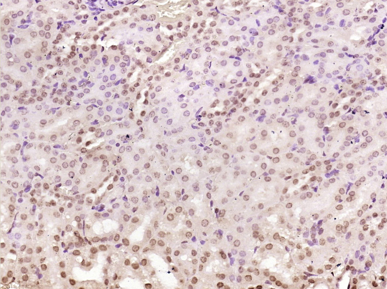
Paraformaldehyde-fixed, paraffin embedded (Mouse kidney); Antigen retrieval by boiling in sodium citrate buffer (pH6.0) for 15min; Block endogenous peroxidase by 3% hydrogen peroxide for 20 minutes; Blocking buffer (normal goat serum) at 37°C for 30min; Antibody incubation with (NADPH oxidase 4) Polyclonal Antibody, Unconjugated (bs-1091R) at 1:400 overnight at 4°C, followed by operating according to SP Kit(Rabbit) (sp-0023) instructions and DAB staining.
-
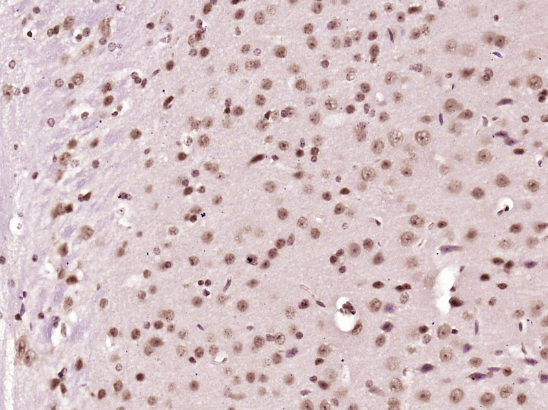
Tissue/cell: rat brain tissue; 4% Paraformaldehyde-fixed and paraffin-embedded;
Antigen retrieval: citrate buffer ( 0.01M, pH 6.0 ), Boiling bathing for 15min; Block endogenous peroxidase by 3% Hydrogen peroxide for 30min; Blocking buffer (normal goat serum,C-0005) at 37℃ for 20 min;
Incubation: Anti-Nox4 Polyclonal Antibody, Unconjugated(bs-1091R) 1:200, overnight at 4°C, followed by conjugation to the secondary antibody(SP-0023) and DAB(C-0010) staining
-
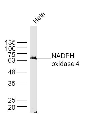
Sample: Hela Cell Lysate at 40 ug
Primary: Anti- NADPH oxidase 4 (bs-1091R) at 1/300 dilution
Secondary: IRDye800CW Goat Anti-Rabbit IgG at 1/20000 dilution
Predicted band size: 64 kD
Observed band size: 64 kD
-
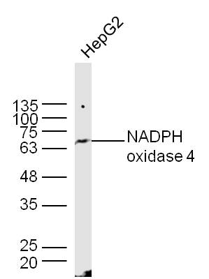
Sample: HepG2 Cell Lysate at 40 ug
Primary: Anti- NADPH oxidase 4(bs-1091R) at 1/300 dilution
Secondary: IRDye800CW Goat Anti-Rabbit IgG at 1/20000 dilution
Predicted band size: 64 kD
Observed band size: 64 kD
-
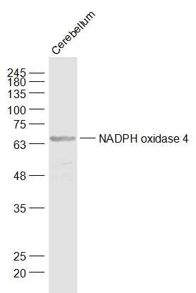
Sample:
Cerebellum (Mouse) Lysate at 40 ug
Primary: Anti-NADPH oxidase 4 (bs-1091R) at 1/1000 dilution
Secondary: IRDye800CW Goat Anti-Rabbit IgG at 1/20000 dilution
Predicted band size: 64 kD
Observed band size: 64 kD
-
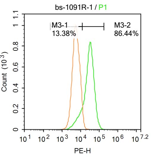
Blank control: Raji.
Primary Antibody (green line): Rabbit Anti-NADPH oxidase 4 antibody (bs-1091R)
Dilution: 1μg /10^6 cells;
Isotype Control Antibody (orange line): Rabbit IgG .
Secondary Antibody : Goat anti-rabbit IgG-PE
Dilution: 1μg /test.
Protocol
The cells were fixed with 4% PFA (10min at room temperature)and then permeabilized with PBST for 20 min at room temperature. The cells were then incubated in 5%BSA to block non-specific protein-protein interactions for 30 min at at room temperature .Cells stained with Primary Antibody for 30 min at room temperature. The secondary antibody used for 40 min at room temperature. Acquisition of 20,000 events was performed.
-
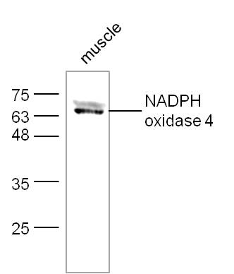
Sample:
muscle (Mouse) Lysate at 40 ug
Primary: Anti-NADPH oxidase 4 (bs-1091R) at 1/300 dilution
Secondary: IRDye800CW Goat Anti-Rabbit IgG at 1/20000 dilution
Predicted band size: 64 kD
Observed band size: 64 kD
-
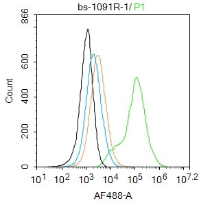
Blank control:293T.
Primary Antibody (green line): Rabbit Anti-NADPH oxidase 4 antibody (bs-1091R)
Dilution: 1μg /10^6 cells;
Isotype Control Antibody (orange line): Rabbit IgG .
Secondary Antibody : Goat anti-rabbit IgG-AF488
Dilution: 1μg /test.
Protocol
The cells were fixed with 4% PFA (10min at room temperature)and then permeabilized with 90% ice-cold methanol for 20 min at -20℃. The cells were then incubated in 5%BSA to block non-specific protein-protein interactions for 30 min at room temperature .Cells stained with Primary Antibody for 30 min at room temperature. The secondary antibody used for 40 min at room temperature. Acquisition of 20,000 events was performed.
-
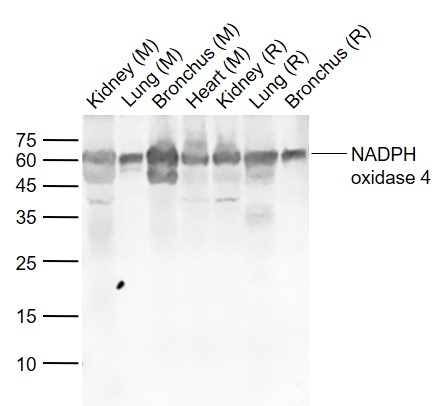
Sample:
Lane 1: Kidney (Mouse) Lysate at 40 ug
Lane 2: Lung (Mouse) Lysate at 40 ug
Lane 3: Bronchus (Mouse) Lysate at 40 ug
Lane 4: Heart (Mouse) Lysate at 40 ug
Lane 5: Kidney (Rat) Lysate at 40 ug
Lane 6: Lung (Rat) Lysate at 40 ug
Lane 7: Bronchus (Rat) Lysate at 40 ug
Primary: Anti-NADPH oxidase 4 (bs-1091R) at 1/1000 dilution
Secondary: IRDye800CW Goat Anti-Rabbit IgG at 1/20000 dilution
Predicted band size: 64 kD
Observed band size: 62 kD
-
Paraformaldehyde-fixed, paraffin embedded (mouse kidney); Antigen retrieval by boiling in sodium citrate buffer (pH6.0) for 15min; Block endogenous peroxidase by 3% hydrogen peroxide for 20 minutes; Blocking buffer (normal goat serum) at 37°C for 30min; Antibody incubation with (NADPH oxidase 4) Polyclonal Antibody, Unconjugated (bs-1091R) at 1:200 overnight at 4°C, followed by operating according to SP Kit(Rabbit) (sp-0023) instructionsand DAB staining.
-
Paraformaldehyde-fixed, paraffin embedded (rat kidney); Antigen retrieval by boiling in sodium citrate buffer (pH6.0) for 15min; Block endogenous peroxidase by 3% hydrogen peroxide for 20 minutes; Blocking buffer (normal goat serum) at 37°C for 30min; Antibody incubation with (NADPH oxidase 4) Polyclonal Antibody, Unconjugated (bs-1091R) at 1:200 overnight at 4°C, followed by operating according to SP Kit(Rabbit) (sp-0023) instructionsand DAB staining.
-
Paraformaldehyde-fixed, paraffin embedded (mouse heart); Antigen retrieval by boiling in sodium citrate buffer (pH6.0) for 15min; Block endogenous peroxidase by 3% hydrogen peroxide for 20 minutes; Blocking buffer (normal goat serum) at 37°C for 30min; Antibody incubation with (NADPH oxidase 4) Polyclonal Antibody, Unconjugated (bs-1091R) at 1:200 overnight at 4°C, followed by operating according to SP Kit(Rabbit) (sp-0023) instructionsand DAB staining.
-
Paraformaldehyde-fixed, paraffin embedded (rat heart); Antigen retrieval by boiling in sodium citrate buffer (pH6.0) for 15min; Block endogenous peroxidase by 3% hydrogen peroxide for 20 minutes; Blocking buffer (normal goat serum) at 37°C for 30min; Antibody incubation with (NADPH oxidase 4) Polyclonal Antibody, Unconjugated (bs-1091R) at 1:200 overnight at 4°C, followed by operating according to SP Kit(Rabbit) (sp-0023) instructionsand DAB staining.
-
Paraformaldehyde-fixed, paraffin embedded (Human kidney ); Antigen retrieval by boiling in sodium citrate buffer (pH6.0) for 15min; Block endogenous peroxidase by 3% hydrogen peroxide for 20 minutes; Blocking buffer (normal goat serum) at 37°C for 30min; Antibody incubation with (NADPH oxidase 4) Polyclonal Antibody, Unconjugated (bs-1091R) at 1:200 overnight at 4°C, followed by operating according to SP Kit(Rabbit) (sp-0023) instructionsand DAB staining.
RRID:AB_10859347
产品名称:Rabbit Anti-NADPH oxidase 4 antibody
别名: KOX 1; KOX; Nox 4; Nox-4; NADPH oxidase 4; RENOX; Kidney oxidase-1; Kidney superoxide-producing NADPH oxidase; Kox-1; NADPH; Nox4; NOX4_HUMAN; Renal NAD(P)H-oxidase; RENOX.
中文名称:NADPH氧化酶4抗体
英文名称:Rabbit Anti-NADPH oxidase 4 antibody
抗体来源: Rabbit
克隆类型:多克隆
细胞定位:细胞浆,细胞膜
性 状:Liquid
亚 型:IgG
纯化方法:affinity purified by Protein A
保存条件:Shipped at 4℃. Store at -20 °C for one year. Avoid repeated freeze/thaw cycles.
免 疫 原:KLH conjugated synthetic peptide derived from human Nox-4
抗原表位:81-180/578
抗原细胞定位:Cytoplasmic
SWISS:Q9JHI8
Gene ID :50507
Human Gene ID:50507
Nox4 is a renal gp91-phox homolog highly expressed at the site of erythropoietin production in the proximal convoluted tubule epithelial cells of the renal cortex. Nox4 is also expressed in fetal tissues, placenta, glioblastoma and vascular cells. Like gp91-phox, the enzymatic activity of Nox4 produces superoxide anions. In vascular cells, the addition of angiotensin II increases Nox4 expression, which suggests a role for Nox-4 in vascular oxidative stress response.
Function:Constitutive NADPH oxidase which generates superoxide intracellularly upon formation of a complex with CYBA/p22phox. Regulates signaling cascades probably through phosphatases inhibition. May function as an oxygen sensor regulating the KCNK3/TASK-1 potass
Subunit:Interacts with, relocalizes and stabilizes CYBA/p22phox. Interacts with TLR4. Interacts with protein disulfide isomerase.
Subcellular Location:Endoplasmic reticulum membrane; Multi-pass membrane protein. Cell junction, focal adhesion. Cell membrane. Note=May localize to plasma membrane and focal adhesions.
Tissue Specificity:EXpressed in brain, in all layers of the cerebellum, in pyramidal cells of the Ammon horn and in Purkinje cells (at protein level). Expressed in osteoclasts, leukocytes, kidney, liver and lung.
Similarity:Contains 1 FAD-binding FR-type domain.
Contains 1 ferric oxidoreductase domain.
Important Note:This product as supplied is intended for research use only, not for use in human, therapeutic or diagnostic applications.
 400-901-9800


 说明书
说明书 联系我们
联系我们 打印此页面
打印此页面 收藏
收藏