欢迎访问博奥森抗体公司官方网站!网站测试中
首页 > 产品 > 一抗 > 产品信息
Rabbit Anti-Tau antibody
多克隆  |   SKU:bs-0157R

说明书

联系我们

打印此页面

收藏

货号:bs-3351R
¥1280
订购号:bs-0157R
¥1098.00-2900.00
货期:现货
Rabbit Anti-Tau antibody
反应物种(预测)

Rabbit

产品应用(已验证)

WB,IHC,FCM

产品应用(可尝试)

IF,ELISA

推荐稀释比例

WB=1:500-2000,Elisa=1:5000-10000,IHC-P=1:100-500,IHC-F=1:100-500,Flow Cyt=1μg/Test,IF=1:100-500,

研究领域

细胞生物,神经生物学,信号转导,细胞凋亡

标签

Array

  • Tissue/cell: Rat brain tissue;4% Paraformaldehyde-fixed and paraffin-embedded;
    Antigen retrieval: citrate buffer ( 0.01M, pH 6.0 ), Boiling bathing for 15min; Blocking buffer (normal goat serum,C-0005) at 37℃ for 20 min;
    Incubation: Anti-MAPT Polyclonal Antibody, Unconjugated(bs-0157R) 1:200, overnight at 4°C; The secondary antibody was Goat Anti-Rabbit IgG, Cy3 conjugated(bs-0295G-Cy3)used at 1:200 dilution for 40 minutes at 37°C. DAPI(5ug/ml,blue,C-0033) was used to stain the cell nuclei
  • Tissue/cell: rat brain tissue; 4% Paraformaldehyde-fixed and paraffin-embedded;
    Antigen retrieval: citrate buffer ( 0.01M, pH 6.0 ), Boiling bathing for 15min; Block endogenous peroxidase by 3% Hydrogen peroxide for 30min; Blocking buffer (normal goat serum,C-0005) at 37℃ for 20 min;
    Incubation: Anti-Tau protein Polyclonal Antibody, Unconjugated(bs-0157R) 1:200, overnight at 4°C, followed by conjugation to the secondary antibody(SP-0023) and DAB(C-0010) staining
  • Blank control (blue line): MCF7 (blue).
    Primary Antibody (green line): Rabbit Anti-Tau antibody (bs-0157R)
    Dilution: 0.2μg /10^6 cells;
    Isotype Control Antibody (orange line): Rabbit IgG .
    Secondary Antibody (white blue line): Goat anti-rabbit IgG-FITC
    Dilution: 1μg /test.
    Protocol
    The cells were fixed with 80% ethanol (Overnight at 4℃) and then permeabilized with 90% ice-cold methanol for 30 min on ice. Cells stained with Primary Antibody for 30 min at room temperature. The cells were then incubated in 1 X PBS/2%BSA/10% goat serum to block non-specific protein-protein interactions followed by the antibody for 15 min at room temperature. The secondary antibody used for 40 min at room temperature. Acquisition of 20,000 events was performed.
  • Positive control: MCF 7 cells(blue)
    (Ice-cold 70% ethanol was added and the cells were incubated for overnight at 4 °C, and then resuspended in ice-cold 90% methanol for 30 min on ice)
    Primary Antibody:Rabbit Anti-Tau antibody (bs-0157R,green)
    Isotype Control Antibody: Rabbit IgG(bs-0295P,orange)
    Secondary Antibody: F(ab’)2 fragment goat anti-rabbit IgG-FITC(White blue)
  • Paraformaldehyde-fixed, paraffin embedded (mouse brain tissue); Antigen retrieval by boiling in sodium citrate buffer (pH6.0) for 15min; Block endogenous peroxidase by 3% hydrogen peroxide for 20 minutes; Blocking buffer (normal goat serum) at 37°C for 30min; Antibody incubation with (Tau) Polyclonal Antibody, Unconjugated (bs-0157R) at 1:200 overnight at 4°C, followed by operating according to SP Kit(Rabbit) (sp-0023) instructionsand DAB staining.
  • Paraformaldehyde-fixed, paraffin embedded (rat brain tissue); Antigen retrieval by boiling in sodium citrate buffer (pH6.0) for 15min; Block endogenous peroxidase by 3% hydrogen peroxide for 20 minutes; Blocking buffer (normal goat serum) at 37°C for 30min; Antibody incubation with (Tau) Polyclonal Antibody, Unconjugated (bs-0157R) at 1:200 overnight at 4°C, followed by operating according to SP Kit(Rabbit) (sp-0023) instructionsand DAB staining.
  • Sample:
    Hippocampus (Rat) Lysate at 40 ug
    Primary: Anti- Tau (bs-0157R) at 1/1000 dilution
    Secondary: IRDye800CW Goat Anti-Rabbit IgG at 1/20000 dilution
    Predicted band size: 52/79 kD
    Observed band size: 52 kD
  • Sample:
    Cerebellum(Mouse) Lysate at 40 ug
    Cerebellum(Rat) Lysate at 40 ug
    BV2(Mouse) Cell Lysate at 40 ug
    A549(Human) Cell Lysate at 40 ug
    RSC96(Rat) CellLysate at 40 ug
    VSC4.1(Rat) Cell Lysate at 40 ug
    Primary: Anti-Tau (bs-0157R) at 1/300 dilution
    Secondary: IRDye800CW Goat Anti-Rabbit IgG at 1/20000 dilution
    Predicted band size: 79 kD
    Observed band size: 52/135 kD
  • Sample:
    Brain (Mouse) Lysate at 40 ug
    Brain (Rat) Lysate at 40 ug
    A549(Human) Cell Lysate at 40 ug
    Primary: Anti-Tau (bs-0157R) at 1/300 dilution
    Secondary: IRDye800CW Goat Anti-Rabbit IgG at 1/20000 dilution
    Predicted band size: 79 kD
    Observed band size: 52/135 kD
  • Sample:
    Lane 1: Cerebrum (Rat) Lysate at 40 ug
    Lane 2: Cerebrum (Mouse) Lysate at 40 ug
    Lane 3: Cerebellum (Rat) Lysate at 40 ug
    Lane 4: Cerebellum (Mouse) Lysate at 40 ug
    Primary: Anti-Tau (bs-0157R) at 1/1000 dilution
    Secondary: IRDye800CW Goat Anti-Rabbit IgG at 1/20000 dilution
    Predicted band size: 50-70 kD
    Observed band size: 50 kD

产品信息

RRID:AB_10855883
产品名称:Rabbit Anti-Tau antibody
别名: MAPT; Microtuble-associted protein Tau; AI413597; AW045860; DDPAC; Disinhibition dementia parkinsonism amyotrophy complex; FLJ31424; FTDP 17; FTDP17; G Protein beta 1 gamma 2 subunit interacting factor 1; G protein beta1/gamma2 subunit interacting factor
中文名称:微管相关蛋白抗体
英文名称:Rabbit Anti-Tau antibody
抗体来源: Rabbit
克隆类型:多克隆
细胞定位:细胞浆,细胞膜
性 状:Liquid
亚 型:IgG
纯化方法:affinity purified by Protein A
保存条件:Shipped at 4℃. Store at -20 °C for one year. Avoid repeated freeze/thaw cycles.

免疫原信息

免 疫 原:KLH conjugated synthetic peptide derived from human Tau
抗原表位:681-758/758
SWISS:P10636
Gene ID :4137
Human Gene ID:4137

产品介绍

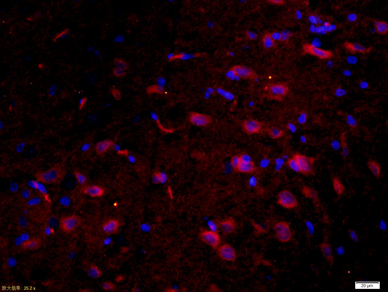
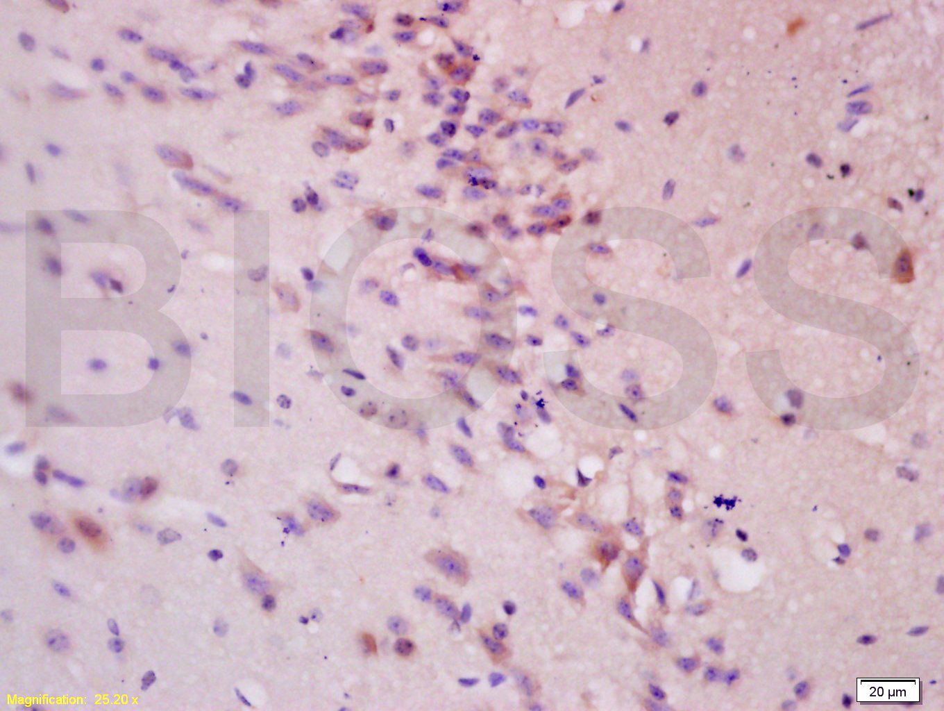
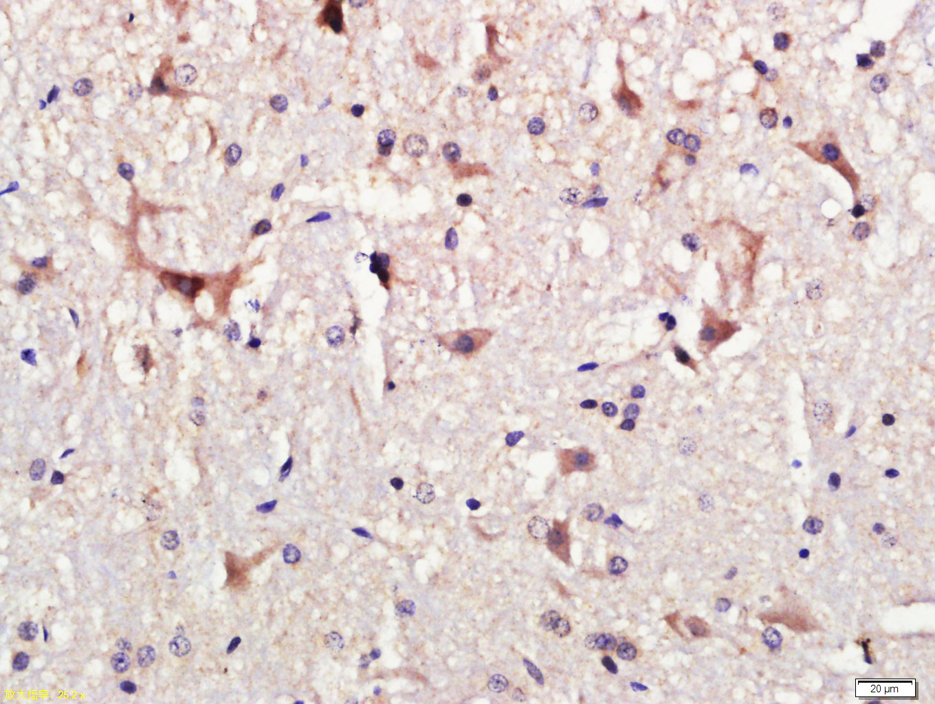
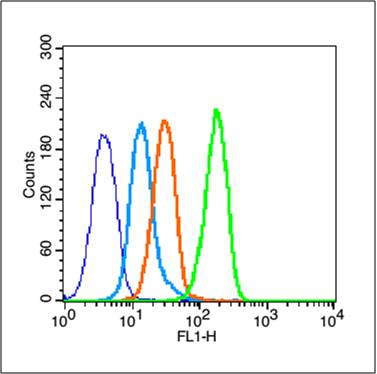
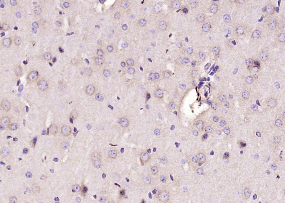
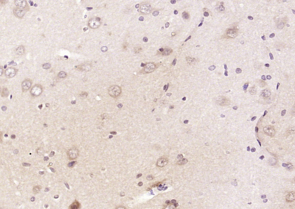
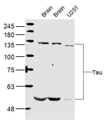
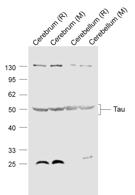
Tau proteins are important Promotes microtubule assembly and stability, and might be involved in the establishment and maintenance of neuronal polarity. The C-terminus binds axonal microtubules while the N-terminus binds neural plasma membrane components, suggesting that tau functions as a linker protein between both. Axonal polarity is predetermined by tau localization (in the neuronal cell) in the domain of the cell body defined by the centrosome. The short isoforms allow plasticity of the cytoskeleton whereas the longer isoforms may preferentially play a role in its stabilization. Tau proteins subcellular located in the axons of neurons, in the cytoso l and in association with plasma membrane components. It expressed in neurons. PNS-tau is expressed in the peripheral nervous system while the others are expressed in the central nervous system.
Function:Promotes microtubule assembly and stability, and might be involved in the establishment and maintenance of neuronal polarity. The C-terminus binds axonal microtubules while the N-terminus binds neural plasma membrane components, suggesting that tau functi
Subunit:Interacts with PSMC2 through SQSTM. Interacts with SQSTM1 when polyubiquitinated. Interacts with FKBP4. Binds to CSNK1D. Interacts with SGK1.
Subcellular Location:Cytoplasm, cytosol. Cell membrane; Peripheral membrane protein; Cytoplasmic side. Cytoplasm, cytoskeleton. Cell projection, axon. Note=Mostly found in the axons of neurons, in the cytosol and in association with plasma membrane components.
Tissue Specificity:Expressed in neurons. Isoform PNS-tau is expressed in the peripheral nervous system while the others are expressed in the central nervous system.
Post-translational modifications:Phosphorylation at serine and threonine residues in S-P or T-P motifs by proline-directed protein kinases (PDPK1: CDK1, CDK5, GSK3, MAPK) (only 2-3 sites per protein in interphase, seven-fold increase in mitosis, and in the form associated with paired hel
DISEASE:Note=In Alzheimer disease, the neuronal cytoskeleton in the brain is progressively disrupted and replaced by tangles of paired helical filaments (PHF) and straight filaments, mainly composed of hyperphosphorylated forms of TAU (PHF-TAU or AD P-TAU). O-Glc
Similarity:Contains 4 Tau/MAP repeats.
Important Note:This product as supplied is intended for research use only, not for use in human, therapeutic or diagnostic applications.

产品订购:sales@bioss.com.cn
邮政编码:101102
综合邮箱:bioss@bioss.com.cn
联系地址:北京市通州区马驹桥镇景盛南四街联东U谷西区2号院67号楼
版权所有 2019-2023 www.bioss.com.cn 北京博奥森生物技术有限公司 京ICP备05066980号-8 京公网安备110107000727号