| Rabbit Anti-CCR7 antibody |
| 反应物种(预测) |
Dog |
| 产品应用(已验证) |
IHC,ICC,FCM |
| 产品应用(可尝试) |
WB,IF,ELISA |
| 推荐稀释比例 |
WB=1:500-2000,Elisa=1:5000-10000,IHC-P=1:100-500,IHC-F=1:100-500,Flow Cyt=1μg/Test,IF=1:100-500,ICC=1:100, |
| 研究领域 |
免疫学,信号转导,G蛋白偶联受体,G蛋白信号, |
| 标签 |
Array |
-
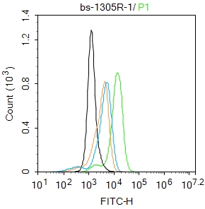
Blank control:THP-1.
Primary Antibody (green line): Rabbit Anti-CCR7 antibody (bs-1305R)
Dilution: 1μg /10^6 cells;
Isotype Control Antibody (orange line): Rabbit IgG .
Secondary Antibody : Goat anti-rabbit IgG-FITC
Dilution: 0.5μg /test.
Protocol
The cells were fixed with 4% PFA (10min at room temperature)and then permeabilized with 0.1% PBST for 20 min at room temperature.The cells were then incubated in 5%BSA to block non-specific protein-protein interactions for 30 min at room temperature .Cells stained with Primary Antibody for 30 min at room temperature. The secondary antibody used for 40 min at room temperature. Acquisition of 20,000 events was performed.
-
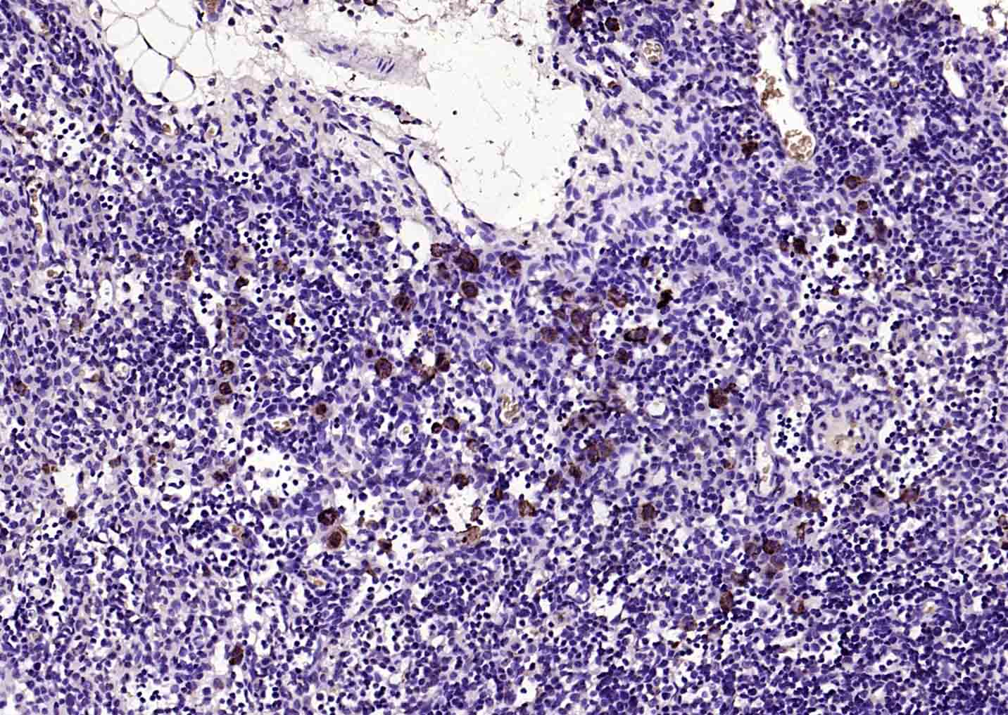
Paraformaldehyde-fixed, paraffin embedded (RAT lymphoid); Antigen retrieval by boiling in sodium citrate buffer (pH6.0) for 15min; Block endogenous peroxidase by 3% hydrogen peroxide for 20 minutes; Blocking buffer (normal goat serum) at 37°C for 30min; Antibody incubation with (CCR7) Polyclonal Antibody, Unconjugated (bs-1305R) at 1:200 overnight at 4°C, followed by operating according to SP Kit(Rabbit) (sp-0023) instructionsand DAB staining.
-
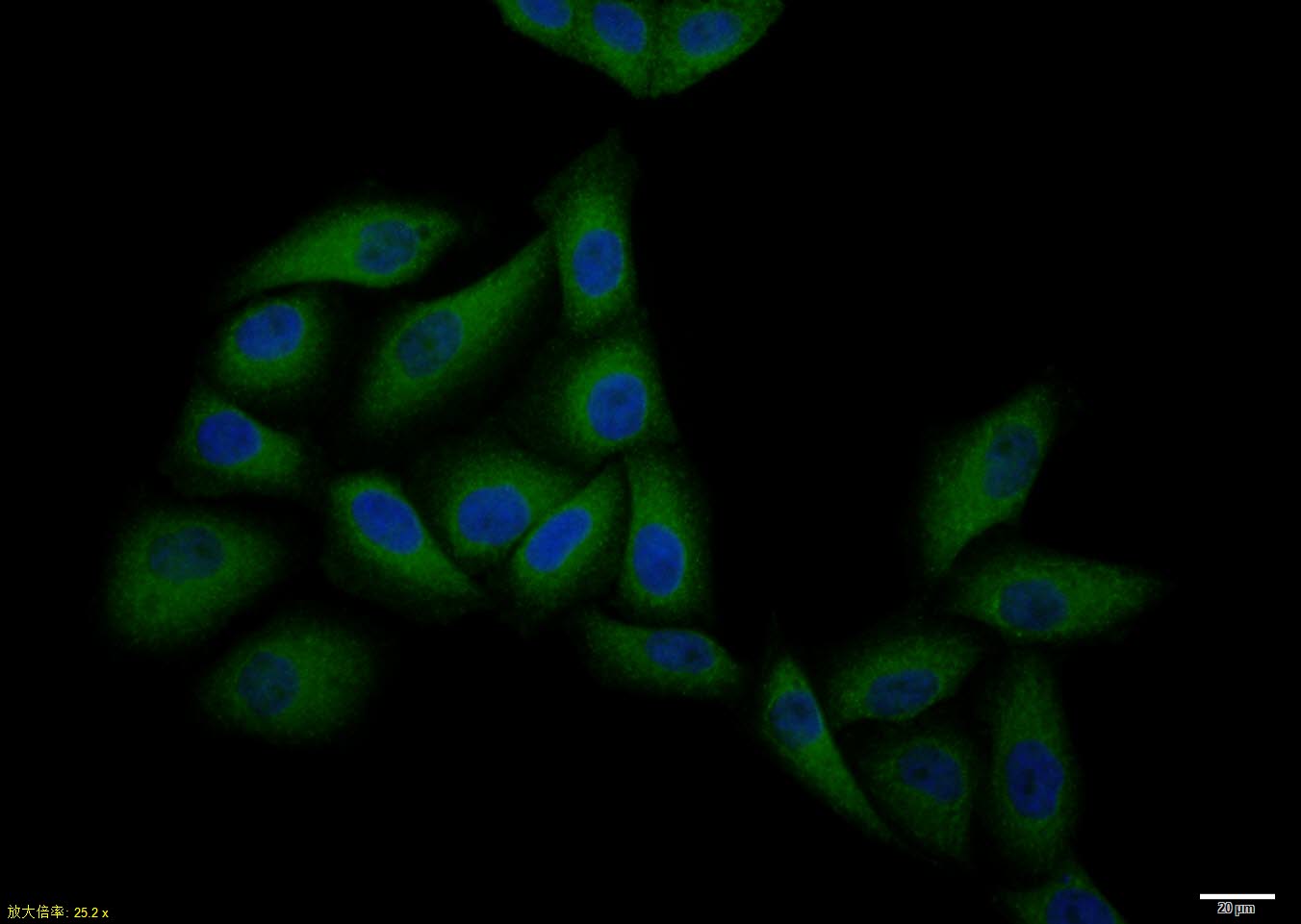
Hela cell; 4% Paraformaldehyde-fixed; Triton X-100 at room temperature for 20 min; Blocking buffer (normal goat serum, C-0005) at 37°C for 20 min; Antibody incubation with (CCR7) polyclonal Antibody, Unconjugated (bs-1305R) 1:100, 90 minutes at 37°C; followed by a conjugated Goat Anti-Rabbit IgG antibody at 37°C for 90 minutes, DAPI (blue, C02-04002) was used to stain the cell nuclei.
-
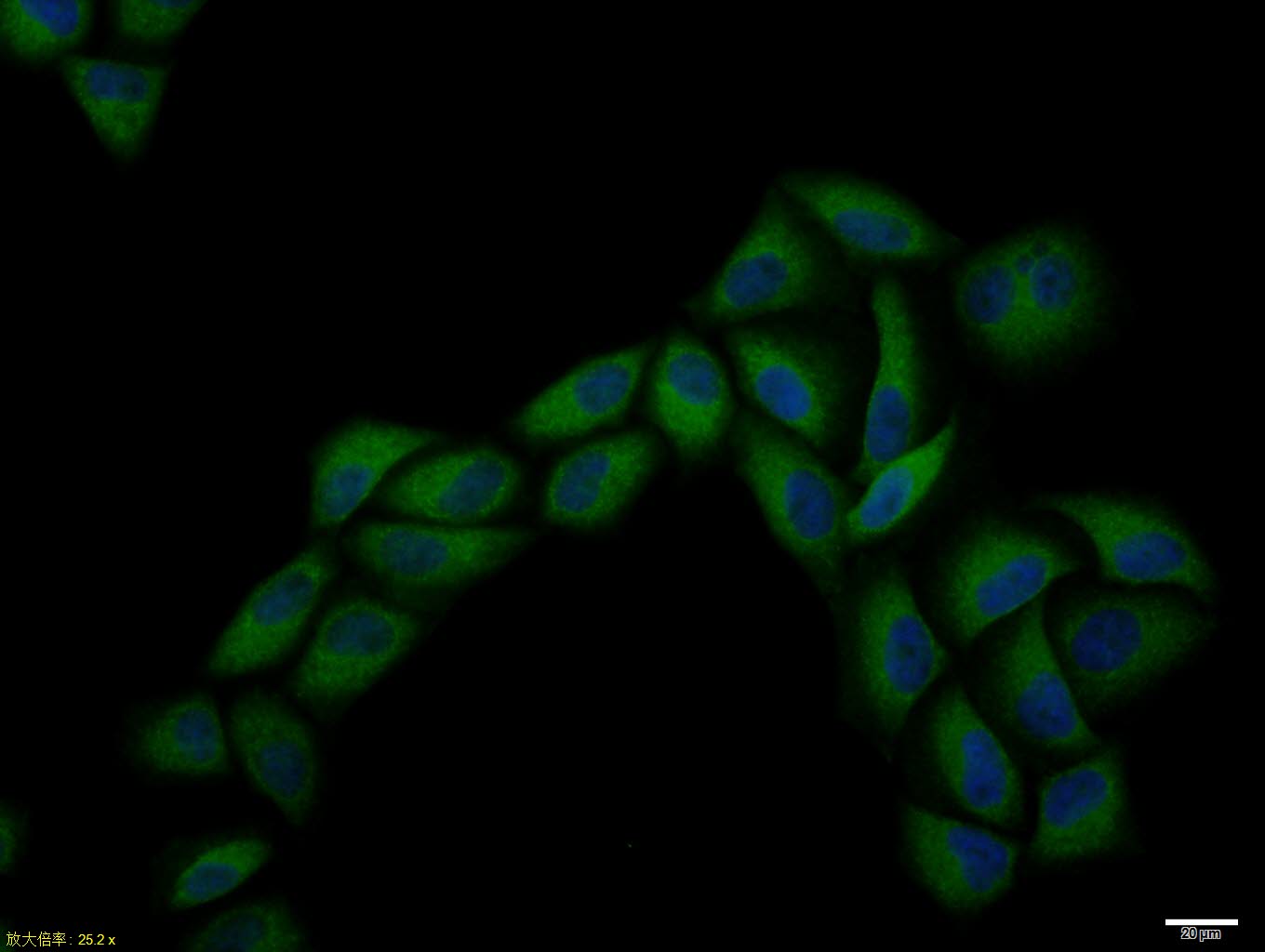
Hela cell; 4% Paraformaldehyde-fixed; Triton X-100 at room temperature for 20 min; Blocking buffer (normal goat serum, C-0005) at 37°C for 20 min; Antibody incubation with (CCR7) polyclonal Antibody, Unconjugated (bs-1305R) 1:100, 90 minutes at 37°C; followed by a conjugated Goat Anti-Rabbit IgG antibody at 37°C for 90 minutes, DAPI (blue, C02-04002) was used to stain the cell nuclei.
-
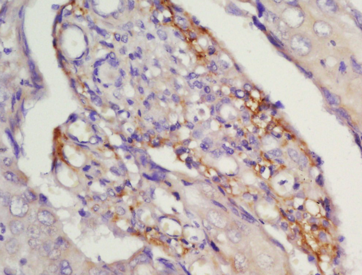
Tissue/cell: human laryngocarcinoma; 4% Paraformaldehyde-fixed and paraffin-embedded;
Antigen retrieval: citrate buffer ( 0.01M, pH 6.0 ), Boiling bathing for 15min; Block endogenous peroxidase by 3% Hydrogen peroxide for 30min; Blocking buffer (normal goat serum,C-0005) at 37℃ for 20 min;
Incubation: Anti-CCR7 Polyclonal Antibody, Unconjugated(bs-1305R) 1:200, overnight at 4°C, followed by conjugation to the secondary antibody(SP-0023) and DAB(C-0010) staining
-
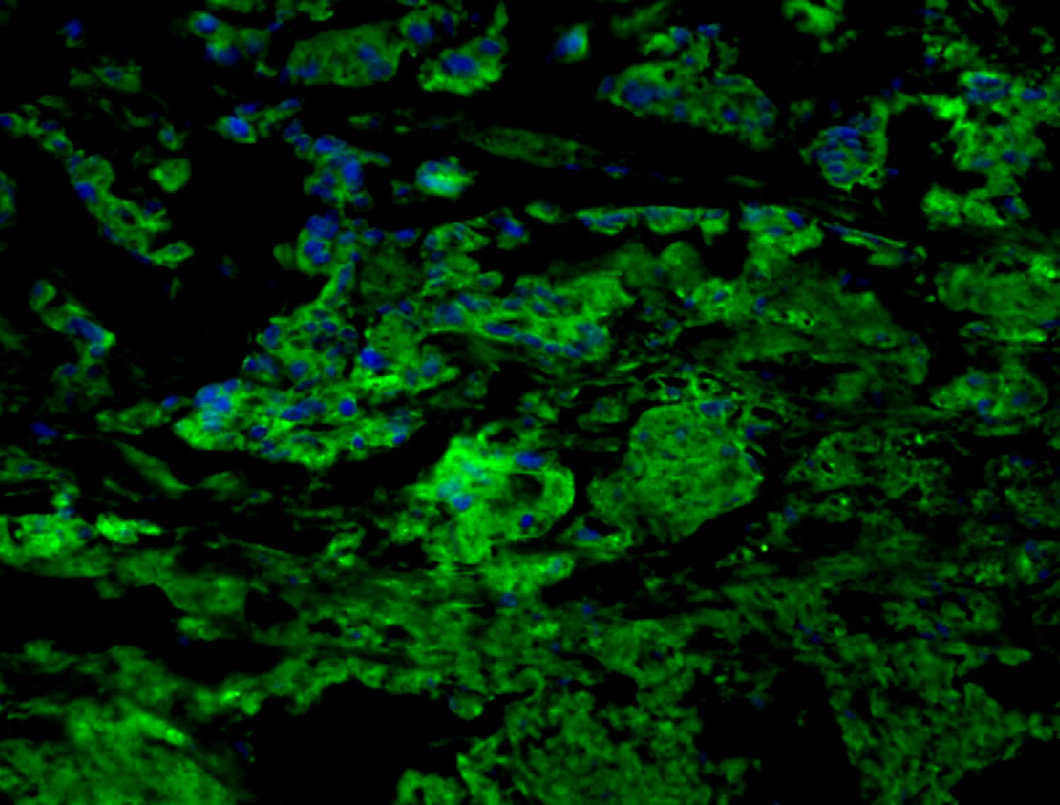
Tissue/cell: human gastric tissue;4% Paraformaldehyde-fixed and paraffin-embedded;
Antigen retrieval: citrate buffer ( 0.01M, pH 6.0 ), Boiling bathing for 15min; Blocking buffer (normal goat serum,C-0005) at 37℃ for 20 min;
Incubation: Anti-CCR7 Polyclonal Antibody, Unconjugated(bs-1305R) 1:200, overnight at 4°C; The secondary antibody was Goat Anti-Rabbit IgG, FITC conjugated(bs-0295G-FITC)used at 1:200 dilution for 40 minutes at 37°C. DAPI(5ug/ml,blue,C-0033) was used to stain the cell nuclei
-
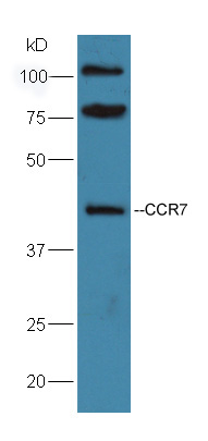
Sample:Raji Cell Lysate at 40 ug
Primary: Anti-CCR7 (bs-1305R) at 1:300 dilution;
Secondary: HRP conjugated Goat-Anti-Rabbit IgG(bs-0295G-HRP) at 1: 5000 dilution;
Predicted band size : 42 kD
Observed band size : 42 kD
-
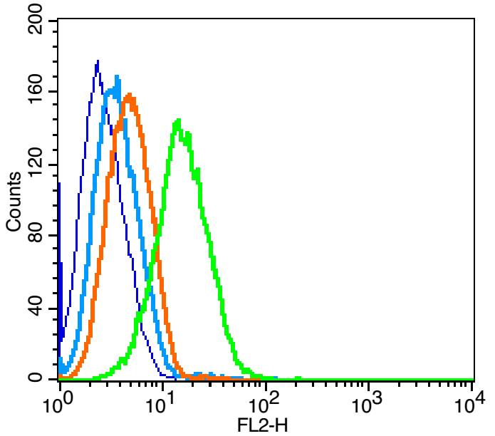
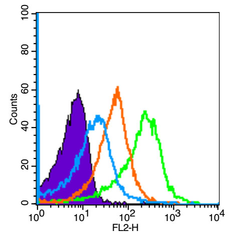
Blank control: Raji(blue).
Primary Antibody:Rabbit Anti-CCR7 antibody(bs-1305R), Dilution: 1μg in 100 μL 1X PBS containing 0.5% BSA;
Isotype Control Antibody: Rabbit IgG(orange) ,used under the same conditions );
Secondary Antibody: Goat anti-rabbit IgG-PE(white blue), Dilution: 1:200 in 1 X PBS containing 0.5% BSA.
Protocol
The cells were fixed with 2% paraformaldehyde (10 min) . Primary antibody (bs-1305R, 1μg /1x10^6 cells) were incubated for 30 min on the ice, followed by 1 X PBS containing 0.5% BSA + 10% goat serum (15 min) to block non-specific protein-protein interactions. Then the Goat Anti-rabbit IgG/PE antibody was added into the blocking buffer mentioned above to react with the primary antibody at 1/200 dilution for 30 min on ice. Acquisition of 20,000 events was performed.
-
Blank control (Black line): Mouse spleen(Black).
Primary Antibody (green line): Rabbit Anti-CD4 antibody (bs-1305R-PE)
Dilution: 3μg /10^6 cells;
Isotype Control Antibody (orange line): Rabbit IgG-PE.
Secondary Antibody (white blue line): Goat anti-rabbit IgG-PE
Dilution: 1μg /test.
Protocol
The cells were fixed with 4% PFA (10min at room temperature)and then permeabilized with 90% ice-cold methanol for 20 min at room temperature. The cells were then incubated in 5%BSA to block non-specific protein-protein interactions for 30 min at room temperature .Cells stained with Primary Antibody for 30 min at room temperature.The secondary antibody used for 40 min at room temperature. Acquisition of 20,000 events was performed.
RRID:AB_10885919
产品名称:Rabbit Anti-CCR7 antibody
别名: CCR7_HUMAN; BLR 2; BLR2; C C chemokine receptor type 7; C C CKR 7; CC chemokine receptor 7; CC chemokine receptor type 7; CC CKR 7; CCCKR7; CCR 7; CD 197; CD197; CD197 antigen; CDW197; Chemokine C C motif receptor 7; Chemokine C C receptor 7; Chemokine re
中文名称:细胞表面趋化因子受体7抗体
英文名称:Rabbit Anti-CCR7 antibody
抗体来源: Rabbit
克隆类型:多克隆
细胞定位:细胞膜
性 状:Liquid
亚 型:IgG
纯化方法:affinity purified by Protein A
保存条件:Shipped at 4℃. Store at -20 °C for one year. Avoid repeated freeze/thaw cycles.
免 疫 原:KLH conjugated synthetic peptide derived from human CCR7
抗原表位:25-59/379
抗原细胞定位:Extracellular
SWISS:P47774
Gene ID :1236
Human Gene ID:1236
The protein encoded by this gene is a member of the G protein-coupled receptor family. This receptor was identified as a gene induced by the Epstein-Barr virus (EBV), and is thought to be a mediator of EBV effects on B lymphocytes. This receptor is expressed in various lymphoid tissues and activates B and T lymphocytes. It has been shown to control the migration of memory T cells to inflamed tissues, as well as stimulate dendritic cell maturation. The chemokine (C-C motif) ligand 19 (CCL19/ECL) has been reported to be a specific ligand of this receptor. [provided by RefSeq, Jul 2008]
Function:Receptor for the MIP-3-beta chemokine. Probable mediator of EBV effects on B-lymphocytes or of normal lymphocyte functions.
Subcellular Location:Cell membrane; Multi-pass membrane protein.
Tissue Specificity:Expressed in various lymphoid tissues and activated B- and T-lymphocytes, strongly up-regulated in B-cells infected with Epstein-Barr virus and T-cells infected with herpesvirus 6 or 7.
Similarity:Belongs to the G-protein coupled receptor 1 family.
Important Note:This product as supplied is intended for research use only, not for use in human, therapeutic or diagnostic applications.
 400-901-9800


 说明书
说明书 联系我们
联系我们 打印此页面
打印此页面 收藏
收藏