欢迎访问博奥森抗体公司官方网站!网站测试中
首页 > 产品 > 一抗 > 产品信息
Mouse Anti-AKT1 antibody
多克隆  |   SKU:bs-0115M

说明书

联系我们

打印此页面

收藏

货号:bs-3351R
¥1280
订购号:bs-0115M
¥1098.00-2900.00
货期:现货
Mouse Anti-AKT1 antibody
反应物种(预测)

Chicken,Dog,Pig,Cow,Rabbit,Sheep

产品应用(已验证)

WB,IHC,ICC

产品应用(可尝试)

IF,ELISA

推荐稀释比例

WB=1:500-2000,Elisa=1:5000-10000,IHC-P=1:100-500,IHC-F=1:100-500,IF=1:100-500,ICC=1:100,

研究领域

肿瘤,细胞生物,神经生物学,信号转导,细胞凋亡,激酶和磷酸酶

标签

Array

  • Tissue/cell: Hela cell; 4% Paraformaldehyde-fixed; Triton X-100 at room temperature for 20 min; Blocking buffer (normal goat serum, C-0005) at 37°C for 20 min; Antibody incubation with (MAKT1) polyclonal Antibody, Unconjugated (bs-0115M) 1:100, 90 minutes at 37°C; followed by a conjugated Goat Anti-Mouse IgG-CY3 antibody at 37°C for 90 minutes, DAPI (blue, C02-04002) was used to stain the cell nuclei.
  • Tissue/cell: rat brain tissue; 4% Paraformaldehyde-fixed and paraffin-embedded;
    Antigen retrieval: citrate buffer ( 0.01M, pH 6.0 ), Boiling bathing for 15min; Block endogenous peroxidase by 3% Hydrogen peroxide for 30min; Blocking buffer (normal goat serum,C-0005) at 37∩ for 20 min;
    Incubation: Anti-PKB Polyclonal Antibody, Unconjugated(bs-0115M) 1:200, overnight at 4∑C, followed by conjugation to the secondary antibody(SP-0024) and DAB(C-0010) staining
  • Sample: Placenta (Mouse) Lysate at 30 ug
    Primary: Anti- AKT1 (bsm-0115M) at 1/300 dilution
    Secondary: IRDye800CW Goat Anti-Mouse IgG at 1/20000 dilution
    Predicted band size: 56 kD
    Observed band size: 56 kD
  • Paraformaldehyde-fixed, paraffin embedded (human memmery cancer); Antigen retrieval by boiling in sodium citrate buffer (pH6.0) for 15min; Block endogenous peroxidase by 3% hydrogen peroxide for 20 minutes; Blocking buffer (normal goat serum) at 37°C for 30min; Antibody incubation with (AKT1) Polyclonal Antibody, Unconjugated (bs-0115R) at 1:500 overnight at 4°C, followed by a conjugated secondary (sp-0023) for 20 minutes and DAB staining.
  • Sample:
    Lung (Mouse) Lysate at 40 ug
    Primary: Anti-AKT1 (bs-0115M) at 1/300 dilution
    Secondary: IRDye800CW Goat Anti-Rabbit IgG at 1/20000 dilution
    Predicted band size: 56 kD
    Observed band size: 56 kD
  • Sample: Hela Cell (Human) Lysate at 40 ug
    Primary: Anti-AKT1 (bs-0115M) at 1/300 dilution
    Secondary: IRDye800CW Goat Anti-Mouse IgG at 1/20000 dilution
    Predicted band size: 56 kD
    Observed band size: 60 kD
  • Sample:
    NIH/3T3(Mouse) Cell Lysate at 30 ug
    MCF-7(Human) Cell Lysate at 30 ug
    A549(Human) Cell Lysate at 30 ug
    Primary: Anti-AKT1 (bs-0115M) at 1/1000 dilution
    Secondary: IRDye800CW Goat Anti-Mouse IgG at 1/20000 dilution
    Predicted band size: 60 kD
    Observed band size: 60 kD
  • Tissue/cell: Hela cell; 4% Paraformaldehyde-fixed; Triton X-100 at room temperature for 20 min; Blocking buffer (normal goat serum, C-0005) at 37°C for 20 min; Antibody incubation with (MAKT1) polyclonal Antibody, Unconjugated (bs-0115M) 1:100, 90 minutes at 37°C; followed by a conjugated Goat Anti-Mouse IgG-CY3 antibody at 37°C for 90 minutes, DAPI (blue, C02-04002) was used to stain the cell nuclei.
  • Paraformaldehyde-fixed, paraffin embedded (rat lung); Antigen retrieval by boiling in sodium citrate buffer (pH6.0) for 15min; Block endogenous peroxidase by 3% hydrogen peroxide for 20 minutes; Blocking buffer (normal goat serum) at 37°C for 30min; Antibody incubation with (AKT1) Monoclonal Antibody, Unconjugated (bs-0115M) at 1:200 overnight at 4°C, followed by operating according to SP Kit(Mouse)(sp-0024) instructionsand DAB staining.
  • Paraformaldehyde-fixed, paraffin embedded (mouse liver); Antigen retrieval by boiling in sodium citrate buffer (pH6.0) for 15min; Block endogenous peroxidase by 3% hydrogen peroxide for 20 minutes; Blocking buffer (normal goat serum) at 37°C for 30min; Antibody incubation with (AKT1) Polyclonal Antibody, Unconjugated (bs-0115M) at 1:200 overnight at 4°C, followed by operating according to SP Kit(Rabbit) (sp-0024) instructionsand DAB staining.
  • Tissue/cell:MCF7 cell; 4% Paraformaldehyde-fixed; Triton X-100 at room temperature for 20 min; Blocking buffer (normal goat serum, C-0005) at 37°C for 20 min; Antibody incubation with (AKT1) polyclonal Antibody, Unconjugated (bs-0115M) 1:100, 90 minutes at 37°C; followed by a CY3 conjugated Goat Anti-Mouse IgG antibody at 37°C for 90 minutes, DAPI (blue, C02-04002) was used to stain the cell nuclei.
  • Tissue/cell:MCF7 cell; 4% Paraformaldehyde-fixed; Triton X-100 at room temperature for 20 min; Blocking buffer (normal goat serum, C-0005) at 37°C for 20 min; Antibody incubation with (AKT1) polyclonal Antibody, Unconjugated (bs-0115M) 1:100, 90 minutes at 37°C; followed by a CY3 conjugated Goat Anti-Mouse IgG antibody at 37°C for 90 minutes, DAPI (blue, C02-04002) was used to stain the cell nuclei.
  • Paraformaldehyde-fixed, paraffin embedded (Mouse brain); Antigen retrieval by boiling in sodium citrate buffer (pH6.0) for 15min; Block endogenous peroxidase by 3% hydrogen peroxide for 20 minutes; Blocking buffer (normal goat serum) at 37°C for 30min; Antibody incubation with (AKT1) Monoclonal Antibody, Unconjugated (bs-0115M) at 1:400 overnight at 4°C, followed by operating according to SP Kit(Mouse) (sp-0024) instructionsand DAB staining.

产品信息

RRID:AB_10886461
产品名称:Mouse Anti-AKT1 antibody
别名: AKT 1; AKT; AKT-1; AKT1_HUMAN; C AKT; cAKT; MGC9965; MGC99656; Oncogene AKT1; PKB; PKB alpha; PKB-ALPHA; PRKBA; Protein Kinase B Alpha; Protein kinase B; Proto-oncogene c-Akt; RAC Alpha; RAC alpha serine/threonine protein kinase; RAC; RAC PK Alpha; Rac pr
中文名称:蛋白激酶B抗体
英文名称:Mouse Anti-AKT1 antibody
抗体来源: Mouse
克隆类型:多克隆
细胞定位:细胞核,细胞浆,细胞膜
性 状:Liquid
亚 型:IgG
纯化方法:affinity purified by Protein A
保存条件:Shipped at 4℃. Store at -20 °C for one year. Avoid repeated freeze/thaw cycles.

免疫原信息

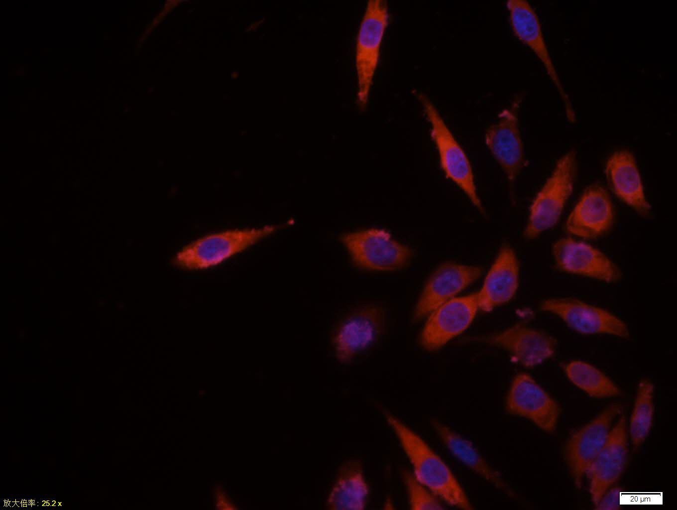
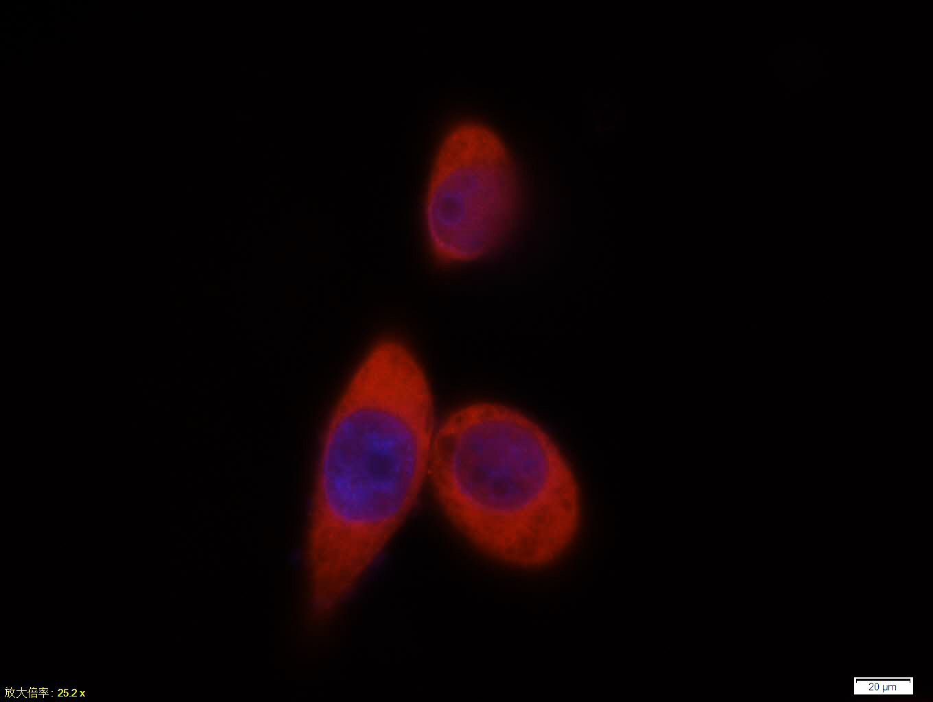
免 疫 原:KLH conjugated synthetic peptide derived from human AKT-1
抗原表位:401-479/479
SWISS:P31749
Gene ID :207
Human Gene ID:207

产品介绍

This gene encodes one of the three members of the human AKT serine-threonine protein kinase family which are often referred to as protein kinase B alpha, beta, and gamma. These highly similar AKT proteins all have an N-terminal pleckstrin homology domain, a serine/threonine-specific kinase domain and a C-terminal regulatory domain. These proteins are phosphorylated by phosphoinositide 3-kinase (PI3K). AKT/PI3K forms a key component of many signalling pathways that involve the binding of membrane-bound ligands such as receptor tyrosine kinases, G-protein coupled receptors, and integrin-linked kinase. These AKT proteins therefore regulate a wide variety of cellular functions including cell proliferation, survival, metabolism, and angiogenesis in both normal and malignant cells. AKT proteins are recruited to the cell membrane by phosphatidylinositol 3,4,5-trisphosphate (PIP3) after phosphorylation of phosphatidylinositol 4,5-bisphosphate (PIP2) by PI3K. Subsequent phosphorylation of both threonine residue 308 and serine residue 473 is required for full activation of the AKT1 protein encoded by this gene. Phosphorylation of additional residues also occurs, for example, in response to insulin growth factor-1 and epidermal growth factor. Protein phosphatases act as negative regulators of AKT proteins by dephosphorylating AKT or PIP3. The PI3K/AKT signalling pathway is crucial for tumor cell survival. Survival factors can suppress apoptosis in a transcription-independent manner by activating AKT1 which then phosphorylates and inactivates components of the apoptotic machinery. AKT proteins also participate in the mammalian target of rapamycin (mTOR) signalling pathway which controls the assembly of the eukaryotic translation initiation factor 4F (eIF4E) complex and this pathway, in addition to responding to extracellular signals from growth factors and cytokines, is disregulated in many cancers. Mutations in this gene are associated with multiple types of cancer and excessive tissue growth including Proteus syndrome and Cowden syndrome 6, and breast, colorectal, and ovarian cancers. Multiple alternatively spliced transcript variants have been found for this gene. [provided by RefSeq, Jul 2020]
Function:AKT1 is one of 3 closely related serine/threonine-protein kinases (AKT1, AKT2 and AKT3) called the AKT kinase, and which regulate many processes including metabolism, proliferation, cell survival, growth and angiogenesis. This is mediated through serine a
Subunit:Interacts (via the C-terminus) with CCDC88A (via its C-terminus). Interacts with GRB10; the interaction leads to GRB10 phosphorylation thus promoting YWHAE-binding. Interacts with AGAP2 (isoform 2/PIKE-A); the interaction occurs in the presence of guanine
Subcellular Location:Cytoplasm. Nucleus. Cell membrane. Note=Nucleus after activation by integrin-linked protein kinase 1 (ILK1). Nuclear translocation is enhanced by interaction with TCL1A. Phosphorylation on Tyr-176 by TNK2 results in its localization to the cell membrane w
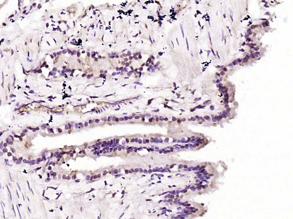
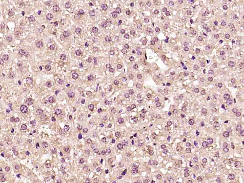
Tissue Specificity:Expressed in prostate cancer and levels increase from the normal to the malignant state (at protein level). Expressed in all human cell types so far analyzed. The Tyr-176 phosphorylated form shows a significant increase in expression in breast cancers dur
Post-translational modifications:O-GlcNAcylation at Thr-305 and Thr-312 inhibits activating phosphorylation at Thr-308 via disrupting the interaction between AKT1 and PDPK1. O-GlcNAcylation at Ser-473 also probably interferes with phosphorylation at this site.
Phosphorylation on Thr-3
DISEASE:Defects in AKT1 are a cause of susceptibility to breast cancer (BC) [MIM:114480]. A common malignancy originating from breast epithelial tissue. Breast neoplasms can be distinguished by their histologic pattern. Invasive ductal carcinoma is by far the mos
Similarity:Belongs to the protein kinase superfamily. AGC Ser/Thr protein kinase family. RAC subfamily.
Contains 1 AGC-kinase C-terminal domain.
Contains 1 PH domain.
Contains 1 protein kinase domain.
Important Note:This product as supplied is intended for research use only, not for use in human, therapeutic or diagnostic applications.

产品订购:sales@bioss.com.cn
邮政编码:101102
综合邮箱:bioss@bioss.com.cn
联系地址:北京市通州区马驹桥镇景盛南四街联东U谷西区2号院67号楼
版权所有 2019-2023 www.bioss.com.cn 北京博奥森生物技术有限公司 京ICP备05066980号-8 京公网安备110107000727号