| Rabbit Anti-F-Actin antibody |
| 反应物种(预测) |
Pig,Cow,Sheep |
| 产品应用(已验证) |
WB,IHC,FCM |
| 产品应用(可尝试) |
ELISA |
| 推荐稀释比例 |
WB=1:1000-5000,Elisa=1:5000-10000,IHC-P=1:100-500,Flow Cyt=3μg /test, |
| 研究领域 |
免疫学,细胞骨架, |
| 标签 |
Array |
-
Paraformaldehyde-fixed, paraffin embedded (rat stomach); Antigen retrieval by boiling in sodium citrate buffer (pH6.0) for 15min; Block endogenous peroxidase by 3% hydrogen peroxide for 20 minutes; Blocking buffer (normal goat serum) at 37°C for 30min; Antibody incubation with (F-Actin) Polyclonal Antibody, Unconjugated (bs-1571R) at 1:200 overnight at 4°C, followed by operating according to SP Kit(Rabbit) (sp-0023) instructionsand DAB staining.
-
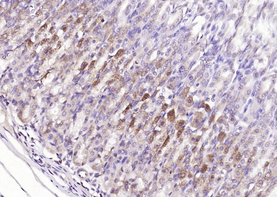
Paraformaldehyde-fixed, paraffin embedded (rat skeletal muscle); Antigen retrieval by boiling in sodium citrate buffer (pH6.0) for 15min; Block endogenous peroxidase by 3% hydrogen peroxide for 20 minutes; Blocking buffer (normal goat serum) at 37°C for 30min; Antibody incubation with (F-Actin) Polyclonal Antibody, Unconjugated (bs-1571R) at 1:200 overnight at 4°C, followed by operating according to SP Kit(Rabbit) (sp-0023) instructionsand DAB staining.
-
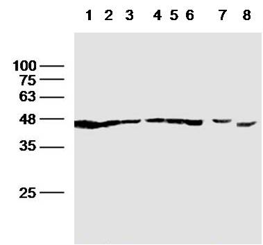
Sample:
Lane1:Hela (Human) Whole Cell Lysate at 30 ug
Lane2:Jurkat (Human) Whole Cell Lysate at 30 ug
Lane3:293T (Human) Whole Cell Lysate at 30 ug
Lane4:Hepg2 (Human) Whole Cell Lysate at 30 ug
Lane5:A431 (Human) Whole Cell Lysate at 30 ug
Lane6:A549 (Human) Whole Cell Lysate at 30 ug
Lane7:H9C2 (Rat) Whole Cell Lysate at 30 ug
Lane8:Testis (Mouse) Tissue Lysate at 30 ug
Primary: Anti-F-Actin(bs-1571R)at 1/300 dilution
Secondary: IRDye800CW Goat Anti-Rabbit IgG at 1/20000 dilution
Predicted band size: 42 kD
Observed band size: 42 kD
-
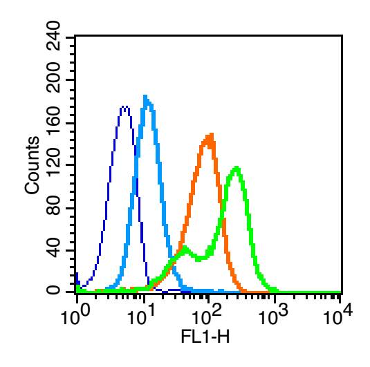
Blank control (blue line): Hela (blue).
Primary Antibody (green line): Rabbit Anti- F-Actin antibody (bs-1571R)
Dilution: 3μg /10^6 cells;
Isotype Control Antibody (orange line): Rabbit IgG .
Secondary Antibody (white blue line): Goat anti-rabbit IgG-PE
Dilution: 1μg /test.
Protocol
The cells were fixed with 80% methanol (5 min at -20℃) and then permeabilized with 0.1% PBS-Tween for 20 min at room temperature . Cells stained with Primary Antibody for 30 min at room temperature. The cells were then incubated in 1 X PBS/2%BSA/10% goat serum to block non-specific protein-protein interactions followed by the antibody for 15 min at room temperature. The secondary antibody used for 40 min at room temperature. Acquisition of 20,000 events was performed.
RRID:AB_10859354
产品名称:Rabbit Anti-F-Actin antibody
别名: f actin; Filamentous actin; F-actin capping protein alpha subunit; CapZ alpha-1; CAZA1_HUMAN; CAPZA1.
中文名称:纤维状肌动蛋白抗体
英文名称:Rabbit Anti-F-Actin antibody
抗体来源: Rabbit
克隆类型:多克隆
细胞定位:细胞浆
性 状:Liquid
亚 型:IgG
纯化方法:affinity purified by Protein A
保存条件:Shipped at 4℃. Store at -20 °C for one year. Avoid repeated freeze/thaw cycles.
免 疫 原:Synthetic MAP peptide derived from human beta-Actin
抗原表位:1-50/374
SWISS:P60709
Gene ID :60
Human Gene ID:60
In vertebrates, three main groups of actin isoforms, alpha, beta, and gamma have been identified. The alpha actins, found in muscletissues, are a major constituent of the contractile apparatus. The beta and gamma actins coexist in most cell types as components of the cytoskeleton, and as mediators of internal cell motility. Individual subunits of microfilaments are known as globular actin (G-actin). G-actin subunits assemble into long filamentous polymers called F-actin.
Important Note:This product as supplied is intended for research use only, not for use in human, therapeutic or diagnostic applications.
 400-901-9800


 说明书
说明书 联系我们
联系我们 打印此页面
打印此页面 收藏
收藏