欢迎访问博奥森抗体公司官方网站!网站测试中
首页 > 产品 > 一抗 > 产品信息
Rabbit Anti-SCF antibody
多克隆  |   SKU:bs-0545R

说明书

联系我们

打印此页面

收藏

货号:bs-3351R
¥1280
订购号:bs-0545R
¥1098.00-2900.00
货期:现货
Rabbit Anti-SCF antibody
产品应用(已验证)

WB,IHC,ICC,FCM

产品应用(可尝试)

IF,ELISA

推荐稀释比例

WB=1:500-2000,Elisa=1:5000-10000,IHC-P=1:100-500,IHC-F=1:100-500,Flow Cyt=1ug/Test,IF=1:100-500,ICC=1:100,

研究领域

细胞生物,干细胞,生长因子和激素,细胞膜蛋白,

标签

Array

  • Tissue/cell: rat spleen tissue;4% Paraformaldehyde-fixed and paraffin-embedded;
    Antigen retrieval: citrate buffer ( 0.01M, pH 6.0 ), Boiling bathing for 15min; Blocking buffer (normal goat serum,C-0005) at 37℃ for 20 min;
    Incubation: Anti-SCF Polyclonal Antibody, Unconjugated(bs-0999R) 1:200, overnight at 4°C; The secondary antibody was Goat Anti-Rabbit IgG, PE conjugated(bs-0295G-PE)used at 1:200 dilution for 40 minutes at 37°C. DAPI(5ug/ml,blue,C-0033) was used to stain the cell nuclei
  • Blank control (Black line): U87MG (Black).
    Primary Antibody (green line): Rabbit Anti-SCF antibody (bs-0545R)
    Dilution: 1μg /10^6 cells;
    Isotype Control Antibody (orange line): Rabbit IgG.
    Secondary Antibody (white blue line): Goat anti-rabbit IgG-PE
    Dilution: 1μg /test.
    Protocol
    The cells were fixed with 4% PFA (10min at room temperature)and then were incubated in 5%BSA to block non-specific protein-protein interactions for 30 min at room temperature .Cells stained with Primary Antibody for 30 min at room temperature.The secondary antibody used for 40 min at room temperature. Acquisition of 20,000 events was performed.
  • Tissue/cell: rat spleen tissue; 4% Paraformaldehyde-fixed and paraffin-embedded;
    Antigen retrieval: citrate buffer ( 0.01M, pH 6.0 ), Boiling bathing for 15min; Block endogenous peroxidase by 3% Hydrogen peroxide for 30min; Blocking buffer (normal goat serum,C-0005) at 37℃ for 20 min;
    Incubation: Anti-SCF Polyclonal Antibody, Unconjugated(bs-0545R) 1:200, overnight at 4°C, followed by conjugation to the secondary antibody(SP-0023) and DAB(C-0010) staining
  • Tissue/cell: rat kidney tissue; 4% Paraformaldehyde-fixed and paraffin-embedded;
    Antigen retrieval: citrate buffer ( 0.01M, pH 6.0 ), Boiling bathing for 15min; Block endogenous peroxidase by 3% Hydrogen peroxide for 30min; Blocking buffer (normal goat serum,C-0005) at 37℃ for 20 min;
    Incubation: Anti-SCF Polyclonal Antibody, Unconjugated(bs-0545R) 1:200, overnight at 4°C, followed by conjugation to the secondary antibody(SP-0023) and DAB(C-0010) staining
  • Paraformaldehyde-fixed, paraffin embedded (Rat brain); Antigen retrieval by boiling in sodium citrate buffer (pH6.0) for 15min; Block endogenous peroxidase by 3% hydrogen peroxide for 20 minutes; Blocking buffer (normal goat serum) at 37°C for 30min; Antibody incubation with (SCF) Polyclonal Antibody, Unconjugated (bs-0545R) at 1:400 overnight at 4°C, followed by operating according to SP Kit(Rabbit) (sp-0023) instructions and DAB staining.
  • Blank control (Black line): Mouse spleen(Black).
    Primary Antibody (green line): Rabbit Anti-SCF antibody (bs-0545R)
    Dilution: 3μg /10^6 cells;
    Isotype Control Antibody (orange line): Rabbit IgG .
    Secondary Antibody (white blue line): Goat anti-rabbit IgG-AF647
    Dilution: 1μg /test.
    Protocol
    The cells were fixed with 4% PFA (10min at room temperature)and then permeabilized with 90% ice-cold methanol for 20 min at room temperature. The cells were then incubated in 5%BSA to block non-specific protein-protein interactions for 30 min at room temperature .Cells stained with Primary Antibody for 30 min at room temperature. The secondary antibody used for 40 min at room temperature. Acquisition of 10,000 events was performed.
  • Sample:
    Lane 1: Lung (Rat) Lysate at 40 ug
    Lane 2: Spleen (Rat) Lysate at 40 ug
    Lane 3: MCF-7 (Human) Cell Lysate at 30 ug
    Lane 4: Hela (Human) Cell Lysate at 30 ug
    Primary: Anti-SCF (bs-0545R) at 1/1000 dilution
    Secondary: IRDye800CW Goat Anti-Rabbit IgG at 1/20000 dilution
    Predicted band size: 45/31/19 kD
    Observed band size: 45/35 kD
  • Hela cell; 4% Paraformaldehyde-fixed; Ice-cold methanol at -20℃ for 20 min; Blocking buffer (normal goat serum, C-0005) at 37°C for 20 min; Antibody incubation with (SCF) polyclonal Antibody, Unconjugated (bs-0545R) 1:100, 90 minutes at 37°C; followed by a FITC conjugated Goat Anti-Rabbit IgG antibody at 37°C for 90 minutes, DAPI (blue, C02-04002) was used to stain the cell nuclei.

产品信息

RRID:AB_10855650
产品名称:Rabbit Anti-SCF antibody
别名: Stem Cell Factor; C kit ligand; Ckit ligand; DKFZp686F2250; KIT ligand; Kitl; KITLG; KL 1; KL1; Mast cell growth factor; MGF; SF; SHEP7; Steel factor; Stem cell factor precursor; SCF_HUMAN.
中文名称:干细胞生长因子抗体
英文名称:Rabbit Anti-SCF antibody
抗体来源: Rabbit
克隆类型:多克隆
细胞定位:细胞浆,细胞膜,分泌型蛋白
性 状:Liquid
亚 型:IgG
纯化方法:affinity purified by Protein A
保存条件:Shipped at 4℃. Store at -20 °C for one year. Avoid repeated freeze/thaw cycles.

免疫原信息

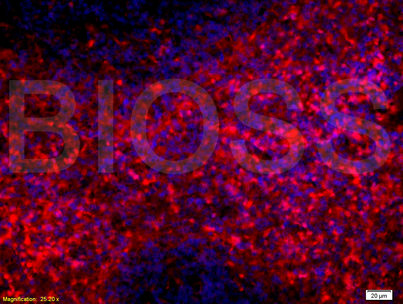
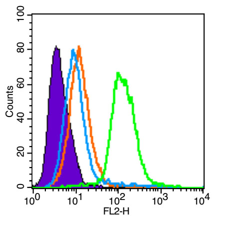
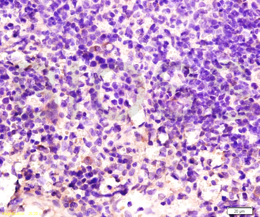
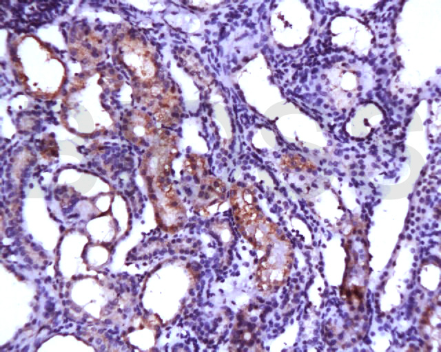
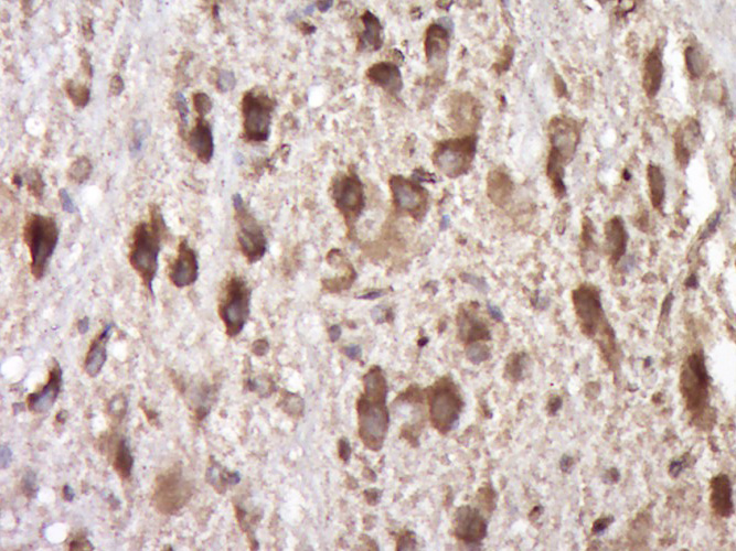
免 疫 原:KLH conjugated synthetic peptide derived from human SCF
SWISS:P21583
Gene ID :4254
Human Gene ID:4254

产品介绍

Stem Cell Factor (SCF), also known as c-Kit ligand (KL), steel factor (SLF) and mast cell growth factor (MGF), is a 30 kDa glycoprotein with broad activities on various tissues, including hematopoietic cells, pigment cells, and primordial germ cells. SCF is secreted by endothelial cells, fibroblasts, and bone marrow stromal cells as a membrane-bound form which may be cleaved to release the soluble form. Both forms are active in promoting colony formation from murine bone marrow cells, but membrane-bound SCF is more effective in promoting hematopoieses in vivo, suggesting a role in cellular interactions between hematopoietic and stromal cells. The soluble form is thought to exist in solution as a noncovalently linked dimer. SCF is structurally related to M-CSF (CSF-1) and Flt-3/Flk-2 Ligand (FL) with all three sharing a similar size, existence of transmembrane and soluble forms, four conserved cysteines, and alternative splicing exon locations, but they share little sequence homology. SCF alone is a modest colony stimulating factor. However, in the presence of other cytokines such as EPO, TPO, GM-CSF, G-CSF, M-CSF, IL-3, and IL-7, SCF is a potent costimulant that works synergistically to increase the size of myeloid, erythroid or lymphoid lineage colonies without influencing the lineage differentiation of the progenitors.
Subunit:Homodimer, non-covalently linked (Probable). Heterotetramer with KIT, binding two KIT molecules; thereby mediates KIT dimerization and subsequent activation by autophosphorylation.
Subcellular Location:Isoform 1: Cell membrane; Single-pass type I membrane protein. Isoform 2: Cell membrane; Single-pass type I membrane protein. Cytoplasm, cytoskeleton. Soluble KIT ligand: Secreted.
Similarity:Belongs to the SCF family.
Important Note:This product as supplied is intended for research use only, not for use in human, therapeutic or diagnostic applications.

产品订购:sales@bioss.com.cn
邮政编码:101102
综合邮箱:bioss@bioss.com.cn
联系地址:北京市通州区马驹桥镇景盛南四街联东U谷西区2号院67号楼
版权所有 2019-2023 www.bioss.com.cn 北京博奥森生物技术有限公司 京ICP备05066980号-8 京公网安备110107000727号