| Rabbit Anti-VCAM1 antibody |
| 反应物种(预测) |
Rat |
| 产品应用(已验证) |
WB,IHC,FCM |
| 产品应用(可尝试) |
IF,ELISA |
| 推荐稀释比例 |
WB=1:500-2000,Elisa=1:5000-10000,IHC-P=1:100-500,IHC-F=1:100-500,Flow Cyt=1µg/Test,IF=1:100-500, |
| 研究领域 |
肿瘤,心血管,细胞生物,细胞粘附分子,内皮细胞, |
| 标签 |
Array |
-
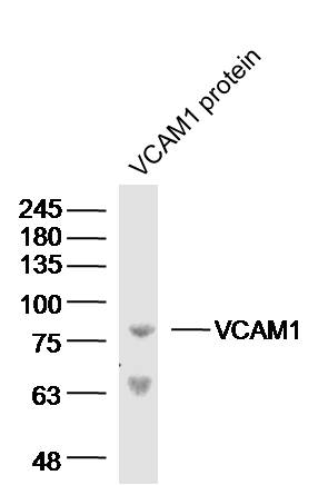
Sample: VCAM1 protein (Human) at 100 ng
Primary: Anti-VCAM-1 (bs-0396R) at 1/300 dilution
Secondary: IRDye800CW Goat Anti-Rabbit IgG at 1/20000 dilution
Predicted band size: 81 kD
Observed band size: 81 kD
-
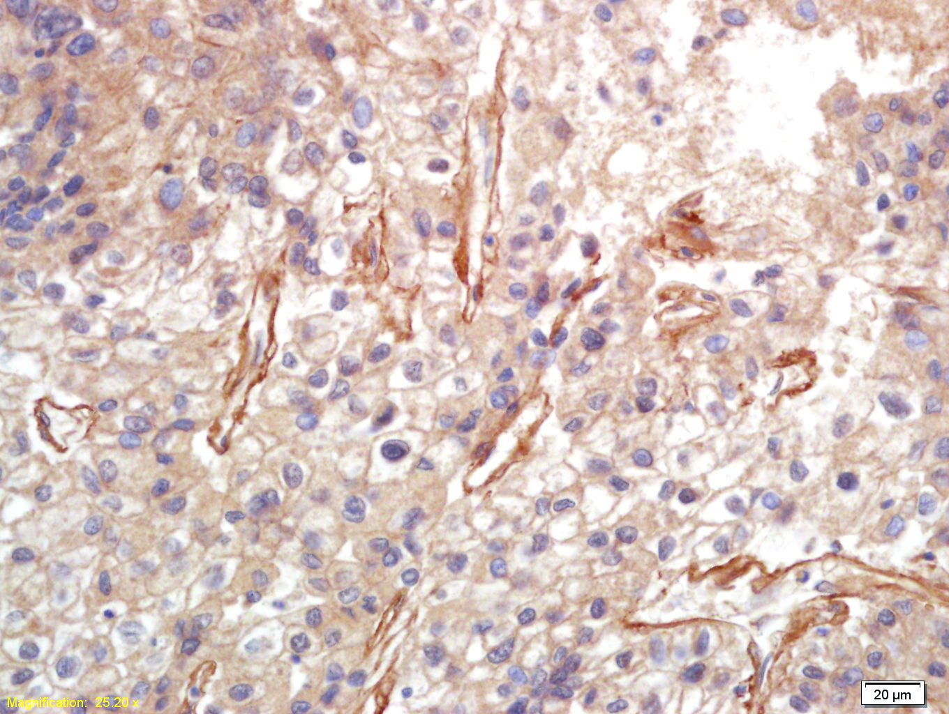
Tissue/cell: human lung carcinoma; 4% Paraformaldehyde-fixed and paraffin-embedded;
Antigen retrieval: citrate buffer ( 0.01M, pH 6.0 ), Boiling bathing for 15min; Block endogenous peroxidase by 3% Hydrogen peroxide for 30min; Blocking buffer (normal goat serum,C-0005) at 37℃ for 20 min;
Incubation: Anti-VCAM-1 Polyclonal Antibody, Unconjugated(bs-0396R) 1:200, overnight at 4°C, followed by conjugation to the secondary antibody(SP-0023) and DAB(C-0010) staining
-
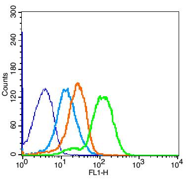
Blank control: Mouse Spleen(blue).
Primary Antibody:Rabbit Anti-VCAM-1 antibody (bs-0396R,Green); Dilution: 1μg in 100 μL 1X PBS containing 0.5% BSA;
Isotype Control Antibody: Rabbit IgG(orange) ,used under the same conditions;
Secondary Antibody: Goat anti-rabbit IgG-FITC(white blue), Dilution: 1:200 in 1 X PBS containing 0.5% BSA.
Protocol
The cells were fixed with 2% paraformaldehyde for 10 min at 37℃. Primary antibody (bs-0396R, 1μg /8x10^5 cells) were incubated for 30 min at room temperature, followed by 1 X PBS containing 0.5% BSA + 1 0% goat serum (1 hour) to block non-specific protein-protein interactions. Then the Goat Anti-rabbit IgG/FITC antibody was added into the blocking buffer mentioned above to react with the primary antibody at 1/200 dilution for 40 min at room temperature. Acquisition of 20,000 events was performed.
-
Sample:
Lane 1: Siha (Human) Cell Lysate at 30 ug
Lane 2: Huvec (Human) Cell Lysate at 30 ug
Lane 3: U251 (Human) Cell Lysate at 30 ug
Primary: Anti-VCAM-1 (bs-0396R) at 1/1000 dilution
Secondary: IRDye800CW Goat Anti-Rabbit IgG at 1/20000 dilution
Predicted band size: 110 kD
Observed band size: 110 kD
RRID:AB_10855959
产品名称:Rabbit Anti-VCAM1 antibody
别名: VCAM1; CD 106; CD106; CD106 Antigen; DKFZp779G2333; INCAM 100; L1CAM; MGC99561; Vascular Cell Adhesion Molecule 1; Vascular cell adhesion protein 1; VCAM 1; VCAM-1; INCAM-100; V-CAM 1; VCAM1_HUMAN.
中文名称:血管内皮细胞粘附分子(CD106)抗体
英文名称:Rabbit Anti-VCAM1 antibody
抗体来源: Rabbit
克隆类型:多克隆
细胞定位:细胞膜
性 状:Liquid
亚 型:IgG
纯化方法:affinity purified by Protein A
保存条件:Shipped at 4℃. Store at -20 °C for one year. Avoid repeated freeze/thaw cycles.
免 疫 原:KLH conjugated synthetic peptide derived from mouse VCAM-1
抗原表位:640-739/739
抗原细胞定位:Extracellular
SWISS:P29533
Gene ID :22329
Human Gene ID:7412
VCAM1 is important in cell-cell recognition. Appears to function in leukocyte-endothelial cell adhesion. Interacts with the integrins alpha4 beta1 (beta 1 integrin VLA4) and alpha4 beta7 on leukocytes, and mediates both adhesion and signal transduction. The VCAM1/VLA4 interaction may play a pathophysiologic role both in immune responses and in leukocyte emigration to sites of inflammation. VCAM1 is also expressed by several non endothelial cell types including some macrophages, follicular dendritic cells and bone marrow, stromal cells.
Function:Important in cell-cell recognition. Appears to function in leukocyte-endothelial cell adhesion. Interacts with the beta-1 integrin VLA4 on leukocytes, and mediates both adhesion and signal transduction. The VCAM1/VLA4 interaction may play a pathophysiolog
Subunit:Binds to ECMV-D capsid proteins and acts as a receptor for this virus.
Subcellular Location:Isoform 1: Cell membrane; Single-pass type I membrane protein. Isoform 2: Cell membrane; Lipid-anchor, GPI-anchor.
Tissue Specificity:Expressed on inflamed vascular endothelium, as well as on macrophage-like and dendritic cell types in both normal and inflamed tissue. Expressed in the bone marrow.
Similarity:Contains 7 Ig-like C2-type (immunoglobulin-like) domains.
Important Note:This product as supplied is intended for research use only, not for use in human, therapeutic or diagnostic applications.
 400-901-9800


 说明书
说明书 联系我们
联系我们 打印此页面
打印此页面 收藏
收藏